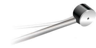
PeriFlux 6000 LDPM/TCPO2
Designed for clinical and preclinical studies, PeriFlux 6000 LDPM leverages laser Doppler perfusion monitoring to provide a range of useful measurements, including blood perfusion.

Laser Doppler for research
Designed for clinical and preclinical studies, PeriFlux 6000 LDPM leverages laser Doppler perfusion monitoring to provide a range of useful measurements, including blood perfusion. By adding a TcpO2 module to PeriFlux 6000 LDPM, precise oxygenation measurements can be gathered, enhancing use in research owing to the complementary nature of LDPM and TcpO2.
In clinical research, PeriFlux 6000 LDPM can be used to, for example, evaluate the effects of drugs or creams on microcirculation, support the development of novel therapies for wound healing and vascular diseases like diabetes, Raynaud’s syndrome, and PAD, and detect underlying conditions such as endothelial dysfunction.
In preclinical studies, PeriFlux 6000 LDPM is suitable for investigating cerebrovascular events like stroke and related conditions.
The technology

Laser Doppler perfusion monitoring is a noninvasive technique designed to measure blood perfusion in the microcirculation — capillaries (nutritive flow), arterioles, venules, and shunting vessels.
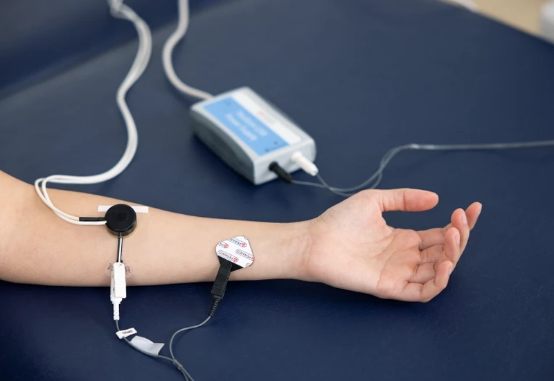
The transmitter in a laser Doppler probe, placed on the skin, emits laser light into the tissue underneath. The light interacts with the tissue, causing it to scatter. Part of the light is absorbed by the tissue or otherwise reflected. When the laser light encounters moving red blood cells, it undergoes a Doppler shift, causing the reflected light to change frequency.
The proportion of Doppler-shifted to non-Doppler-shifted light is directly proportional to the number of red blood cells in the illuminated tissue. The magnitude of the frequency shift is proportional to the velocity of red blood cells. The receiver in the probe detects the reflected light and converts it into an electronic signal. Advanced signal processing in PeriFlux 6000 LDPM converts the data into real-time measurements of microcirculatory blood flow.
How deep measurement occurs in laser Doppler perfusion monitoring primarily depends on the distance between the transmitter and receiver as well as the wavelength of the laser light. Tissue characteristics, such as the structure and density of the capillary beds, pigmentation, and oxygenation, also influence the depth [1].
The proportion of Doppler-shifted to non-Doppler-shifted light is directly proportional to the number of red blood cells in the illuminated tissue. The magnitude of the frequency shift is proportional to the velocity of red blood cells. The receiver in the probe detects the reflected light and converts it into an electronic signal. Advanced signal processing in PeriFlux 6000 LDPM converts the data into real-time measurements of microcirculatory blood flow.
Perimed’s laser Doppler instruments, measured on normal skin, with a 780 nm wavelength laser and a transmitter/receiver separation of 0.25 mm, typically measure depths in the range of 0.3 to 0.5 millimeters. A portion of the signal is influenced by blood flow at depths of 1 mm or more.
In blood-rich organs like the kidney or liver, or in highly scattering tissues like white brain matter, measurement depth is shallower. When blood flow to a region is occluded, the measurement depth may increase since the absence of blood allows more light to pass through the tissue.
Measurements are expressed in perfusion units (PU), which are arbitrary. To ensure comparability of results, probe calibration is essential. Perimed’s probes and instruments are factory-calibrated and we supply a specialized motility standard for accurate calibration verification.
Heat provocation
Perimed Probe 457 is a thermostatic laser Doppler probe designed to measure blood perfusion precisely. It incorporates a heating element that allows for controlled warming of the skin and capture of microvascular blood flow data under different thermal conditions.
This feature is particularly useful for studies on conditions like Raynaud’s phenomenon, diabetes, and other microcirculatory disorders. Together with PeriFlux 6000 LDPM, probe 457 ensures high-quality and reproducible data collection.
Post-occlusive reactive hyperemia
PORH is a physiological response observed after a period (3-5 minutes) of restricted blood flow to tissue. Once pressure is released, a sudden increase in blood flow occurs, surpassing baseline levels by several hundred percent [2]. This hyperemic response serves to rapidly restore oxygen and nutrients and remove metabolic waste accumulated during occlusion.
PORH is often used as a measure of vascular health and endothelial function. PeriFlux 6000 LDPM can measure the degree and duration of hyperemia, enabling researchers to evaluate the reactivity of blood vessels and the overall health of the vascular system.
LDPM and TcpO2
TcpO2 is a noninvasive method used to measure the partial pressure of oxygen at the skin’s surface. It reflects local tissue oxygenation and capillary blood flow — useful markers in the assessment of tissue hypoxia and wound healing potential.
LDPM and TcpO2 are valuable and complementary tools for assessing wound healing potential but TcpO2 readings can sometimes be difficult to interpret as inflammation or edema can lower values. This is where LDPM comes in.
LDPM with iontophoresis
Iontophoresis is a noninvasive method for delivering drugs or other substances through the skin using a mild electrical current. It works on the principle that like charges repel each other, so when an electrically charged drug is placed on the skin and a low-intensity current is applied, the drug molecules are pushed through the skin into the underlying tissue without requiring needles or injections.
Iontophoresis together with laser Doppler perfusion monitoring enables researchers to assess the effect of a substance on the microcirculation.
Our portfolio includes PeriIont, a useful system for studying endothelial dysfunction, and it is available for PeriFlux 6000 LDPM/TcpO2.
An essential biomarker
Blood perfusion serves as a window into the health of the vascular system and provides a quantitative way to assess tissue health, disease progression, and treatment effectiveness.
Tissue viability and wound healing
Reduced perfusion can lead to delayed wound healing or even tissue necrosis. By measuring perfusion, researchers can assess the efficacy of novel treatments to improve blood flow.
Disease progression
For vascular diseases like peripheral artery disease (PAD) impaired blood perfusion can be an indication of disease progression. Tracking perfusion allows researchers to study how conditions evolve and to evaluate interventions aimed at improving blood flow.
Microvascular function
Perfusion measurements can help detect endothelial dysfunction, a precursor to many vascular diseases. By evaluating microvascular blood flow, researchers can study early signs of systemic diseases before they manifest more severely.
Drug development
Blood perfusion measurements are often used in pharmacological studies to evaluate the impact of drugs on microcirculation. It allows researchers to determine if a drug improves blood flow to targeted tissues, aiding in the development of therapies for conditions like stroke or ischemia.
Cerebrovascular health
In stroke research, monitoring cerebral blood perfusion is crucial for understanding how blood flow changes during ischemic events and how it recovers afterward. This has significant implications for developing treatments to restore normal brain function post-stroke.
Example studies
Raynaud’s phenomenon
RP isa condition characterized by episodes of reduced blood flow to the fingers and toes, often triggered by cold or stress, causing them to turn white or blue. In this study [3], 52 adolescents were assessed at the Department of Pediatrics of the Clinical Centre of the University of Pécs, Hungary (March and April, 2015).
Using a PeriFlux 5000 LDPM, baseline finger blood perfusion measurements were taken followed by incremental heat provocations using the heat functionality of Perimed Probe 457. Microcirculation parameters, such as time to peak response, peak perfusion value, and percentage increase (between baseline and heat provocations) were recorded. The study concluded that altered microvascular response to heat provocation could be an early marker of RP.
Wound healing
In one study [4], LD with heat provocation was shown to complement TcpO2 by providing more detailed information, especially in cases where TcpO2 values were misleading due to acute inflammation. In their study of patients with problematic wounds, significant differences were noted between healthy individuals and those with healing issues when both LD and TcpO2 were used together, strengthening the predictive value for wound healing outcomes.
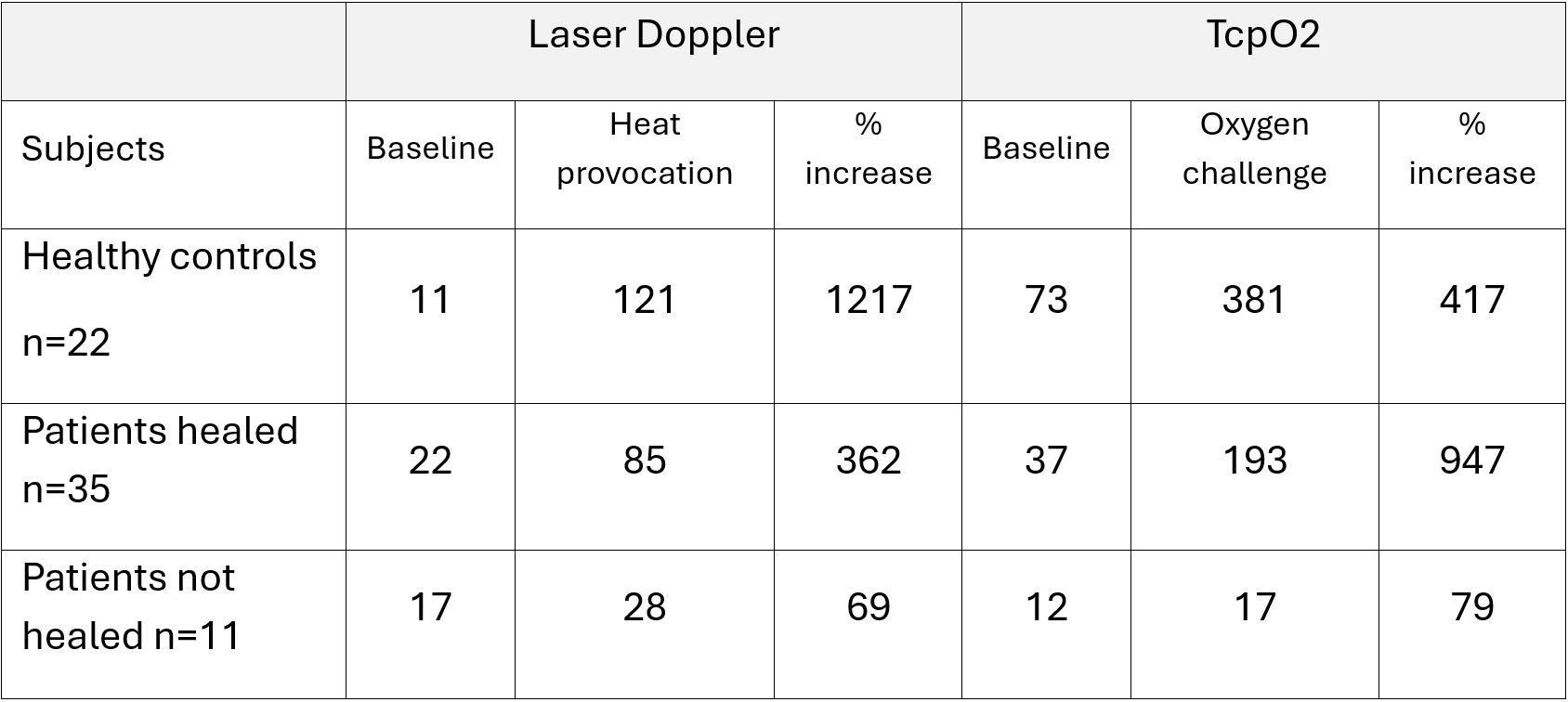
Table 1
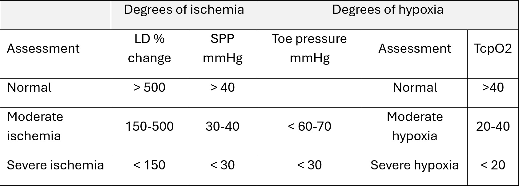
Table 2
Table 2 is a suggested classification system for degrees of ischemia and hypoxia. Perfusion of the local tissue was considered normal with LD percentage increase above 500. Values between 150 and 500 were considered moderately ischemic and values below 150 were considered severely ischemic. In the study [4], LD parameters for healing were LD max> 20 and LD % increase > 150.
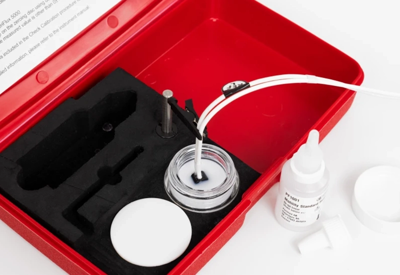
Motility standard
Our Motility Standard is specifically designed for calibrating and verifying Perimed laser Doppler probes, including the 457. It contains a defined volume of colloidal suspension made up of precisely sized polystyrene particles. These particles exhibit well-defined Brownian motion, with a known and constant scatter angle. The magnitude and frequency of the reflected laser Doppler-shifted and non-Doppler-shifted reflected light is also known. Any deviation is automatically resolved by the system to ensure that the laser Doppler measurements of blood perfusion remain precise and reliable across different tests and conditions.
This predictable behavior allows for consistent and reliable performance across different probes and measurement conditions. Probes should be verified at least once every six weeks.
Contact US
Get in touch
For more information about PeriFlux 6000 LDPM with or without TcpO2, fill out the form and we will be in touch with you shortly.
References
- Fredriksson, I., Larsson, M., & Strömberg, T. (2009). Measurement depth and volume in laser Doppler flowmetry. Microvascular research, 78(1), 4–13. https://doi.org/10.1016/j.mvr.2009.02.008
- Sheffield, Paul J. & Smith, Adrianne P.S. & Fife, Caroline E. (2004), Wound Care Practice, Best Publishing Company, ISBN 1-930536-16-X
- Mosdósi, B., Bölcskei, K., & Helyes, Z. (2018). Impairment of microcirculation and vascular responsiveness in adolescents with primary Raynaud phenomenon. Pediatric rheumatology online journal, 16(1), 20. https://doi.org/10.1186/s12969-018-0237-x
- Sheffield PJ, Dietz D, Posey KI, Ziemba TA, Bakken B. Use of transcutaneous oximetry and laser Doppler with local heat provocation to assess patients with problem wounds. In: N M Petri, D Andric, D Ropac (eds), Proceedings, 1st Congress of the Alps-Adria Working Community on Maritime, Undersea, and Hyperbaric Medicine. Split, Croatia: Croatian Maritime, Undersea and Hyperbaric Medical Society of Croatian Medical Association, 2001:341-344.