Exercise TcpO2
Exercise TcpO2, based on DROP (delta from resting oxygen pressure) values, is a noninvasive measurement protocol that provides insight into how well oxygen is delivered to tissue during exertion. It can reveal exercise-induced vascular claudication that may not be apparent at rest or through other measurements such as ABI and TBI.
Physiological principle of Exercise TcpO2
Exercise TcpO2 compares transcutaneous oxygen measured at rest with levels recorded during exertion. Exercise TcpO2 is a provocation test based on the physiological response to exercise, which induces increased oxygen demand.
For a healthy limb, increased oxygen demand from exercise can be met, and TcpO2 remains relatively stable. Arterial disease, however, restricts blood flow, reducing the microcirculation’s ability to deliver oxygen — causing a drop in TcpO2 during exercise.
To enable clinicians to localize restrictions, Exercise TcpO2 typically encompasses DROP measurements at multiple sites — the calf, thigh, and buttocks — on both sides of the body. Recovery rate is also considered valuable for clinical evaluation.
Clinical application
Exercise TcpO2 is particularly useful for assessing patients with suspected PAD when resting tests are inconclusive or unreliable. Current guidelines recommend exercise testing when ABI is normal, but symptoms persist, while several studies demonstrate that exercise TcpO2 improves detection of hemodynamically significant stenoses and is especially valuable in patients with non-compressible arteries or complex comorbidities [1].

Professor Pierre Abraham, one of the pioneers of Exercise TcpO2, provides detailed clinical insights in this post:
How DROP is calculated
To isolate local ischemia from systemic effects, TcpO2 levels are corrected for a reference site. The chest is typically chosen because it is well perfused and generally unaffected by peripheral artery disease. At any point in time (t), DROP is calculated as:
DROP(t) = ΔTcpO2 (limb)(t) – ΔTcpO2 (chest)(t)
For example
Resting TcpO2 for calf = 50mmHg; Resting TcpO2 for chest = 70mmHg
Calf TcpO2 during exercise at time t = 30mmHg; Chest TcpO2 during exercise at time t = 65mmHg
ΔTcpO2 (calf)(t) 50 => 30 = -20 mmHg; ΔTcpO2 (chest)(t) 70 => 65 = -5mmHg
Calf DROP(t): -20 – (-5) = -15mmHg.
DROP is acknowledged and used in clinical research and specialist practice, with a cutoff of ≤- 15 mmHg indicating significant arterial stenosis [2].
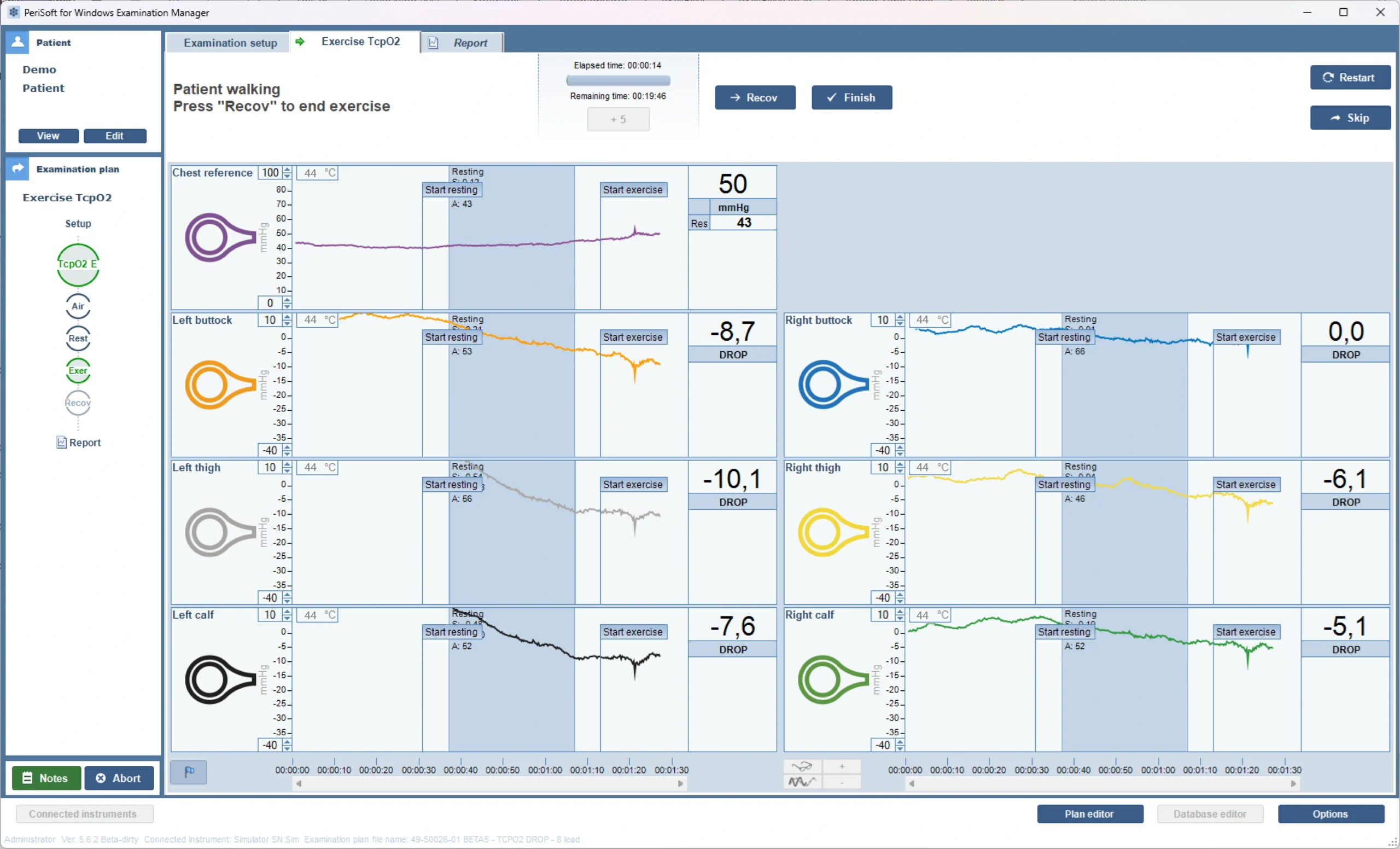
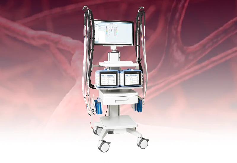
Exercise TcpO2 with PeriFlux 6000
Typically, the patient stands on a treadmill while being prepped. Sites include the buttocks, thighs, and calves on both sides of the body. Preparation is the same as for supine TcpO2. Additional support using split-gauze dressings can help prevent TcpO2 electrodes from moving during the measurement process.
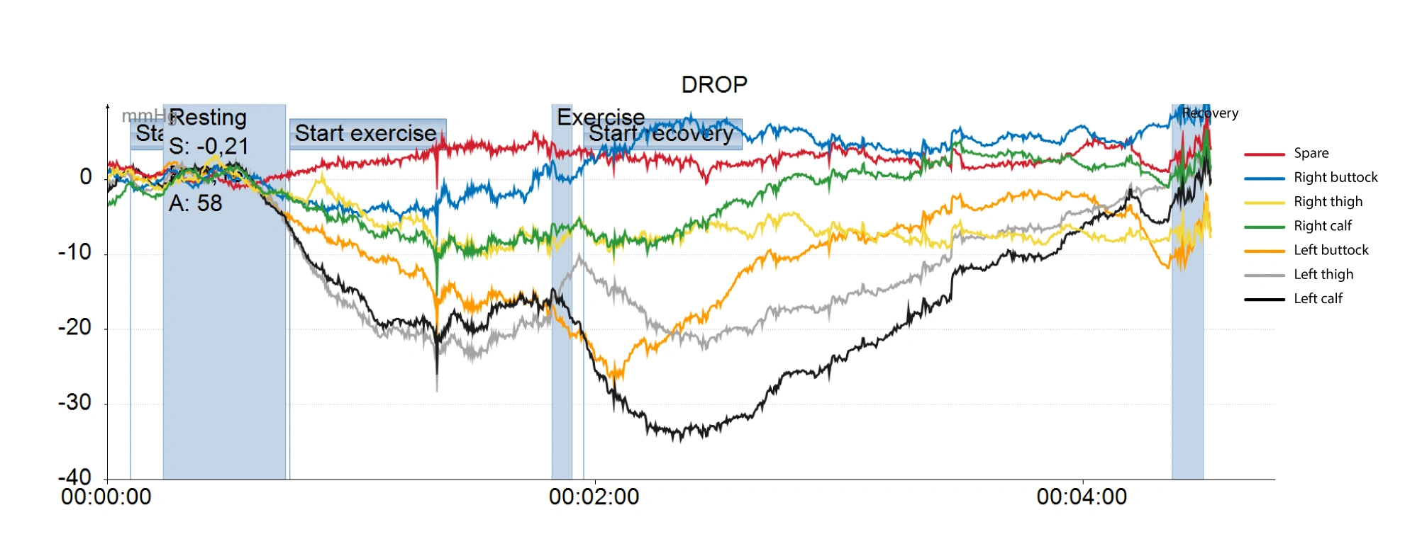
Once the patient is prepped, the Exercise TcpO2 protocol for PeriFlux 6000 visualizes three phases: resting, exercise, and recovery. By default, resting TcpO2 for a limb is calculated as the mean value during the last 30 seconds of the rest period. DROP is calculated for every measurement site in real-time and visualized as color-coded curves.
Curve interpretation
During exercise, a healthy limb will show a relatively stable or rapidly recovering TcpO2. Exercise-induced ischemia is characterized by:
- Delayed recovery
- A significant DROP, below -15mmHg
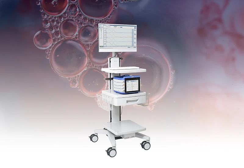
Related products and compatibility
Perimed’s Exercise TcpO2 protocol is available for PeriFlux 6000 Combined System and PeriFlux 6000 TcpO2 System running PSW ExM version 5.6.2 or higher. If you have a PeriFlux 6000 Pressure System, it may be possible to upgrade it to a Combined System to support Exercise TcpO2.
Related products
Contact us
For more information about how to get started with Exercise TcpO2 for PeriFlux 6000, fill out the form, and we will be in touch with you shortly.
References
- Stivalet, O., Paisant, A., Belabbas, D., Le Faucheur, A., Landreau, P., Le Pabic, E., Omarjee, L., & Mahé, G. (2021). Combination of Exercise Testing Criteria to Diagnose Lower Extremity Peripheral Artery Disease. Frontiers in cardiovascular medicine, 8, 759666.
https://doi.org/10.3389/fcvm.2021.759666 - SS08. Exercise Transcutaneous Oxygen Pressure Measurement Is a Reliable Method of Evaluation of Internal Iliac Artery Flow Compromise Sen, Indrani et al. Journal of Vascular Surgery, Volume 67, Issue 6, e87 – e88. DOI: 10.1016/j.jvs.2018.03.086