Chorioallantoic membrane — CAM assay
The CAM assay is a versatile animal model for studying tumor formation, angiogenesis, and metastasis. It is highly reproducible, cost-effective, and has a natural immunodeficiency making it suitable for cell transplantation. The model relies on the creation of a window in the shell of a fertilized chicken egg, which enables researchers to observe the embryo and the formation of blood vessels.
Measuring with PeriCAM PSI HR

PeriCam PSI can be used to measure the (change in) blood perfusion in the angiogenesis assay and detect differences in the efficacy of various pro-angiogenic compounds.
The data enables you to monitor the formation of functional intratumoral blood vessels[1] and validate the effectiveness of anti-angiogenic agents for cancer treatment.
Overcoming data challenges
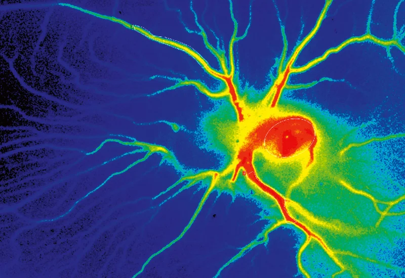
Researchers often use the CAM assay to study angiogenesis, which requires detailed measurements of blood vessel growth, density, and branching patterns. Quantifying these changes using image-based methods can be challenging as varying light conditions and magnification can make it difficult to standardize measurements and achieve reproducible data. PeriCam PSI HR includes automated background compensation and features for setting measurement distance and magnification.
High-resolution imaging is required to capture subtle vascular changes on the CAM. Achieving consistently high image quality without introducing artifacts can be difficult. The translucent nature of the CAM and its sensitivity to light exposure can impact image clarity. PeriCam PSI HR records at a frequency of up to 100 images per second with a max resolution of 10 μm/pixel. Leveraging LSCI. PeriCam PSI HR enables researchers to capture dynamic changes in microcirculation and tissue perfusion with precision.
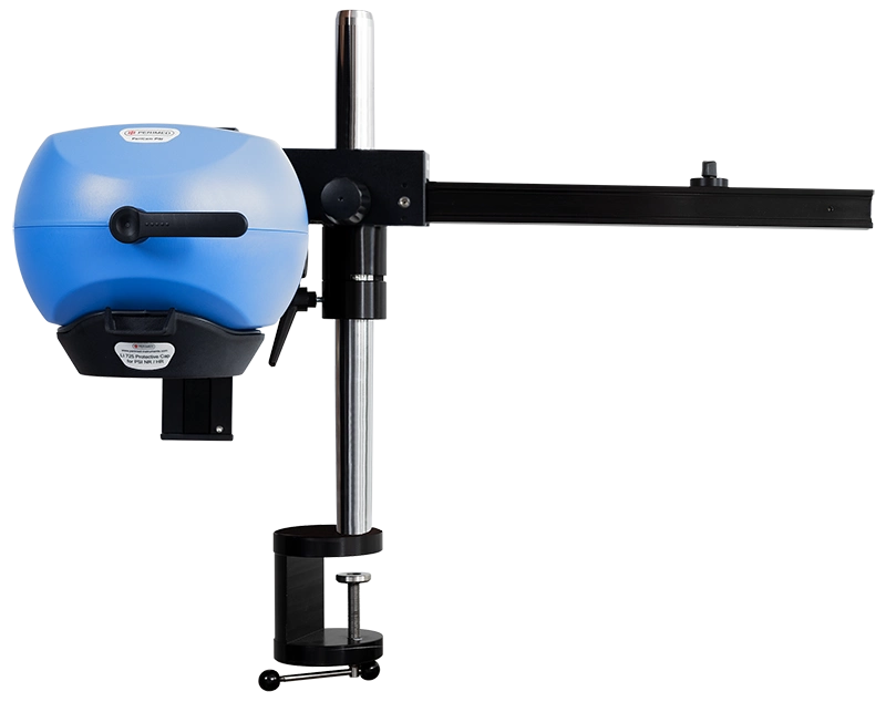
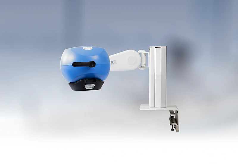
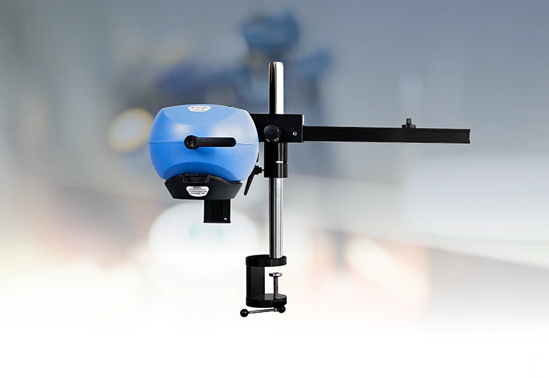
PeriCam PSI HR
PeriCam PSI leverages laser speckle contrast imaging (LSCI) to assess blood perfusion in tissue at the microcirculation level with detail and precision. PeriCam PSI HR (high resolution) works from a fixed distance and is specifically equipped to measure small animals such as rats and mice.
- Rapid data analysis, delivering perfusion graphs, images, and video recordings in real-time.
- Regions of interest (ROIs) and time periods of interest (TOIs) that can be defined and updated before, during, and after recording.
- Frequency of up to ~100 images per second.
- Automated background compensation for robust measurements under varying lighting conditions.
- Suitable for longitudinal studies.
- High-quality arm with good maneuverability to ease positioning, and stability to prevent movement artifacts.
- Wide-angled color camera for documentation of the measurement area and its surroundings, easing post-measurement analysis.
- Non-contact, non-destructive, and noninvasive.
Related products
Contact us
Get in touch
For more information about using PeriCam PSI for a CAM assay or any other preclinical research application, fill out the form, and we will be in touch with you shortly.
References
- Laser speckle contrast analysis (LASCA) technology for the semiquantitative measurement of angiogenesis in in-ovo-tumor-model. Eric Pion, Claudia Asam, Anna-Lena Feder, Oliver Felthaus, Paul I. Heidekrueger,Lukas Prantl, Silke Haerteis, Thiha Aung. 2020, Microvascular Research, Vol. 133, p. 104072.
LASCA and LSCI
Laser speckle contrast imaging (LSCI) is the innovative technology behind PeriCam PSI, offering non-contact, high-resolution perfusion imaging. The technique was first described by J.D. Briers and S. Webster in 1996 and referred to as laser speckle contrast analysis (LASCA). In later years, the term LSCI gained in popularity and is nowadays the more common term. However, LSCI and LASCA both refer to the same technique, providing precise and reliable perfusion data without the need for contrast agents.