PeriCam PSI
Real-time perfusion imaging
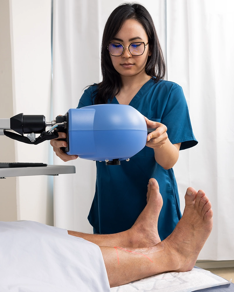
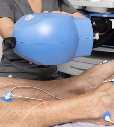
Designed for use in clinical and research settings, PeriCam PSI is a laser-based, non-contact and noninvasive blood perfusion imaging solution.
Leveraging laser speckle contrast imaging (LSCI), PeriCam PSI measures blood perfusion in tissue at the microcirculation level with detail, precision, and in real-time.
Data and images
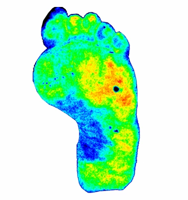
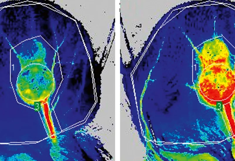
PeriCam PSI transforms blood perfusion in tissue into a real-time data stream of perfusion units. To facilitate interpretation of the data, the application software (PIMSoft) visualizes the measurement area with color coding.
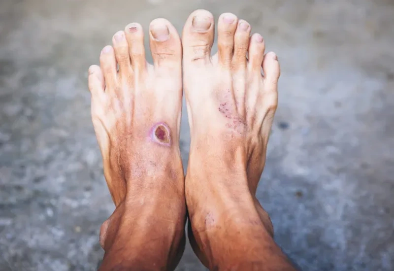
In this perfusion image of a person with a foot ulcer, the color coding indicates lower perfusion in the blue areas compared with the green and yellow ones. Perfusion is low in the wound, with areas of higher perfusion around it.
PeriCam PSI includes a range of color-coding schemes that you can set to suit your practice or established best practices.
Features
- Ease of use — non-contact, noninvasive, and non-destructive.
- Visualization — perfusion graphs and color-coded images and video facilitate data interpretation.
- Robust data — precise, accurate, repeatable, and objective data points to support analysis and longitudinal studies.
- Facilitate analysis — create regions of interest (ROIs) before, during, and after recording and time periods of interest (TOIs) post recording.
- High temporal resolution — with a frequency of up to ~100 images per second.
- Spatial resolution — 10 μm/pixel (PeriCam PSI HR) 33 μm/pixel (PeriCam PSI NR).
- Sensitivity — capable of detecting subtle variations in blood perfusion.
- Continuous autofocus (not all models) — a setup feature to compensate for tissue movements or changes in the instrument’s positioning ensuring every image is sharp and precisely focused on your measurement area.
- Automatic ambient light compensation — to ensure robust measurements under varying lighting conditions.
- High-quality arm — good maneuverability and stability to prevent movement artifacts.
- Wide-angled documentation camera — easing post-measurement analysis.
- Color coding — default color schemes can be adapted to suit your practice and established thresholds.
Noninvasive, non-contact, non-destructive
PeriCam PSI is built on noninvasive and contact-free design principles, making it ideal for measuring perfusion where contact-based measurement techniques are not feasible — such as wounds and surgery.
The technology doesn’t require contrast or tracer agents — reducing the clinical workload, simplifying the measurement process, and lowering the impact on patients and study participants.
PeriCam PSI’s non-contact and noninvasive design makes it a non-destructive tool, helping to preserve the integrity of delicate tissues and minimize impact on the subject.
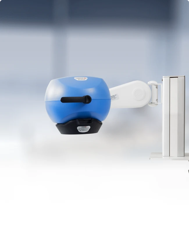
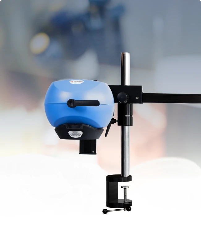
PeriCam PSI models and configurations
To meet the highly diverse needs of clinicians, clinical research, and preclinical scenarios, we’ve designed two models, PeriCam PSI NR and PeriCam PSI HR, with several configurations:
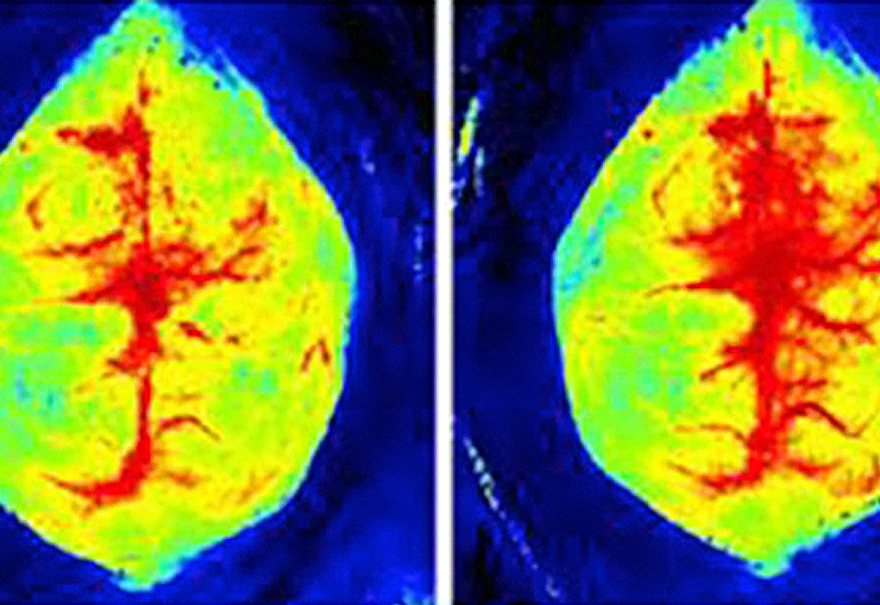
- PeriCam PSI HR — designed for close-up focus, operating at a fixed distance of 13 cm. The camera optics are set to a high magnification to capture details such as small vessels in a mouse brain.
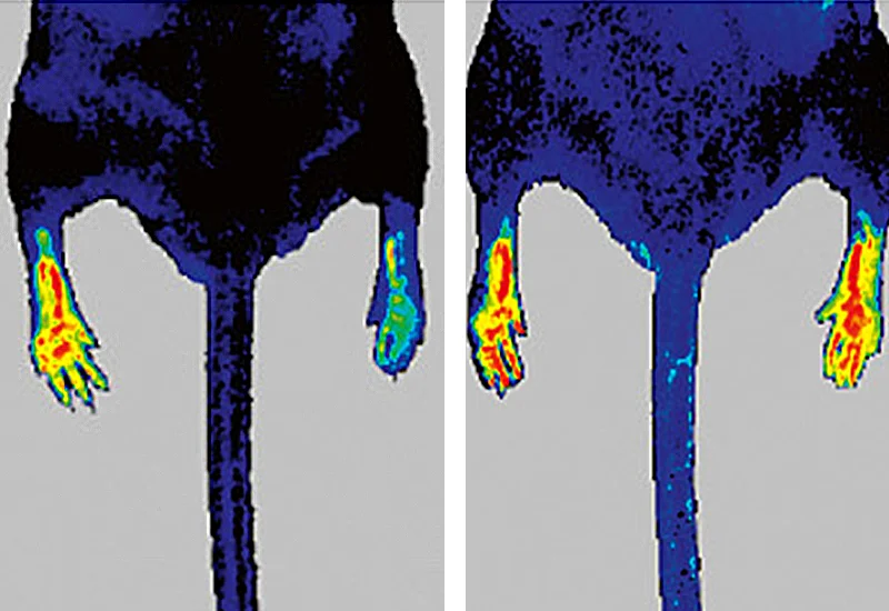
- PeriCam PSI NR — operates at working distances of 13 cm up to 41.5 cm and includes continuous autofocus. The camera optics are set to a viewing angle to capture large areas, such as a human foot.
- Zoom functionality — PeriCam PSI NR and HR can be upgraded with zoom to unlock the camera optics, combining the best of both models. Zoom not only expands the measurement flexibility of your instrument it also broadens its field of application.
- Clinical configuration — includes a PeriCam PSI NR with zoom functionality and high-quality arm mounted on a medical cart. Ships with a high-end PC to ensure performance, a clinical keyboard and mouse, as well as a clinical software package for secure management of patient data.

How it works
Blood perfusion is relative to the volume of moving red blood cells (RBCs) in tissue. To measure perfusion, PeriCam PSI includes several components, including a:
- Near-infrared (NIR) laser to create a speckle pattern on the tissue.
- Positioning laser to create a visible crosshair on the measurement site.
- Highly sensitive camera to record subtle changes in the local contrast between speckles.
- Documentation camera to take ordinary color images of the measurement area, useful for sharing and post-recording analysis.
When you place PeriCam PSI over the measurement area, the NIR laser illuminates the tissue, which creates a speckle pattern.
As the NIR light hits moving red blood cells (RBCs), motion blur occurs in the speckle pattern. The greater the RBC movement, the greater the blur, the lower the contrast between the pixels in the image.
Blood perfusion in the local tissue is inversely proportional to the level of contrast. PeriCam PSI calculates the value as perfusion units in real time.

Some motion blur (on the hand)
Greater motion blur (under the nail)
No motion blur (the table)
Zoom functionality
You can upgrade your PeriCam PSI with zoom to unlock the camera optics, combining the best of PeriCam PSI NR and HR.
- Versatility in clinical and research settings — the adjustable zoom enables you to measure a wide range of tissue types from small vessels in a mouse brain to large areas such as a human foot. Use the same instrument for a range of preclinical and clinical applications such as wound assessment, diabetic foot ulcers, or other conditions where localized perfusion is a key marker.
- High-resolution imaging — the zoom allows for precise visualization of small areas, which is crucial for identifying subtle differences in blood flow, and includes continuous autofocus*, to ensure every image is sharp and precisely focused on your measurement area.
- Enhanced diagnostic capabilities — the ability to zoom in to a critical area, provides clinicians and researchers with a detailed picture of the microvascular health of tissues, potentially leading to more accurate assessments and interventions.
*continuous autofocus is already included in PeriCam PSI NR.
Clinical and research applications
The power of measurement is about:
- Leveraging technology to measure what humans can’t observe.
- Ensuring measurement processes are simple and fast.
- Delivering robust — precise, accurate, repeatable, and objective — measurements.
- Assuring measurements are useful and relevant.
Designed with these principles in mind, our solutions help clinicians and researchers assess and evaluate treatment effectiveness at every intervention — changing outcomes. Here’s an overview of what you can do.
Preclinical research


Color coding
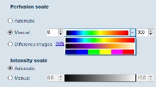
PeriCam PSI uses color coding to represent perfusion levels in a visually intuitive way so that the data is easy to interpret. Different schemes are built into the system. You can also define your own schemes to suit your preferences, the needs of a study, or clinical best practices.
The scale does not affect the underlying data points, only how they are visualized. If you want to compare perfusion images for patient follow-up or in a longitudinal study, for example, they should use the same color-coding scheme. You can, however, always regenerate images from the underlying data.
Default scheme
The default color coding scheme applies to most applications and uses blue, cyan, green, yellow, orange, and red.
- Blue and cyan => low perfusion.
- Green and yellow => medium perfusion.
- Orange and red => high perfusion.
Customizable color schemes
In addition to the default schemes, you can create your own scheme. This is a useful feature if you need to emphasize specific perfusion ranges or mimic an established color convention.
In one study[1], for example, researchers devised a new color palette for PeriCam PSI based on the healing times of a series of burns and a comparison to a similar color scale for laser Doppler imaging. You can set the color scheme of your PeriCam PSI to match the findings of this study.
Contact us
Get in touch
If you would like to know more about PeriCam PSI and how it can help you in your clinical practice or research, fill out the form and we will be in touch with you shortly.
LASCA and LSCI
Laser speckle contrast imaging (LSCI) is the innovative technology behind PeriCam PSI, offering non-contact, high-resolution perfusion imaging. The technique was first described by J.D. Briers and S. Webster in 1996 and referred to as laser speckle contrast analysis (LASCA). In later years, the term LSCI gained in popularity and is nowadays the more common term. However, LSCI and LASCA both refer to the same technique, providing precise and reliable perfusion data without the need for contrast agents.
References
- Annemieke Dijkstra, Goksel Guven, Margriet E. van Baar, Nicole Trommel, Helma W.C. Hofland, T. Martijn Kuijper, Can Ince, C.H. Van der Vlies (2023), Laser speckle contrast imaging, an alternative to laser doppler imaging in clinical practice of burn wound care derivation of a color code, Burns, Volume 49, Issue 8, 2023, Pages 1907-1915. Burns : journal of the International Society for Burn Injuries, 49(8), 1907-1915. https://doi.org/10.1016/j.burns.2023.04.009