Wound healing
Wound healing can be complex and unpredictable. Stakes are high when healing slows or stalls and every wound presents unique challenges. While foundational methods such as visual inspections and touch tests like capillary refill are part of everyday practice, not all wounds respond the same. Conditions like diabetes and peripheral artery disease (PAD) can compromise healing by reducing oxygen and nutrient delivery to the wound site.
Wound healing therapies
None

Wound will heal itself
Conservative

Dressings, offloading, and/or medication
Aided

Advanced treatment like hyperbaric oxygen therapy
Surgery

Advanced treatment like hyperbaric oxygen therapy
The complexity of wound healing often leaves us grappling with important questions:
- How to rapidly assess the potential of a wound to heal.
- Determining appropriate therapy.
- What tools provide objective data to guide care, without increasing workflow complexity.
Beyond visual assessment
Visual and tactile observations provide an essential starting point but don’t always reveal the complete picture. Subtle issues in microcirculation can go unnoticed, delaying the opportunity to intervene.
Technologies like laser speckle contrast imaging (LSCI) and laser Doppler offer deeper insights into tissue health and healing potential beyond visual assessment. When it comes to methods, TcpO2, toe pressure, and SPP are recommended in major international guidelines as an effective tool for wound healing prognosis.
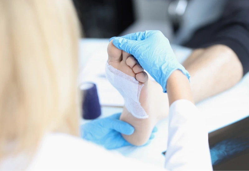
Contact with wounds
Frequent handling of wounds can cause patient discomfort and increase the risk of infection. Solutions that can provide insights about the underlying physiology of the patient in a noninvasive way or without physical contact or contrast agents support a patient-centered and data-driven approach to care.


Workflow efficiency
Solutions should prioritize efficiency — not only the time it takes to measure patients but also the time needed for training and maintenance. Seamless integration with existing patient management systems is critical for sharing results and avoiding time-consuming, error-prone tasks like patient data input.
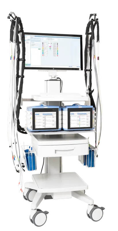
PeriFlux 6000 Combined System
Our combined system offers a wide range of noninvasive macro and microcirculation exams some of which can be conducted simultaneously to reduce time spent in the exam room.
PeriFlux 6000 Combined supports most of the recommended pressure measurements, as well as skin perfusion pressure (SPP) and TcpO2, making it a versatile solution for assessing various wound types across diverse patient conditions and people with amputated toes.
The intuitive user interface is designed to minimize clinical workload by guiding users through the measurement process. All measurements are delivered in a comprehensive report.
Our DICOM/HL7 option connects your PeriFlux 6000 to existing patient management systems for rapid and error-free patient data retrieval and instant report sharing.
With standard workflows for toe pressure and TBI, ankle pressure and ABI, skin perfusion pressure (SPP), TcpO2, and more, PeriFlux 6000 Combined is an ideal tool for wound care centers. For a full list of supported exams and features, please visit the product page.
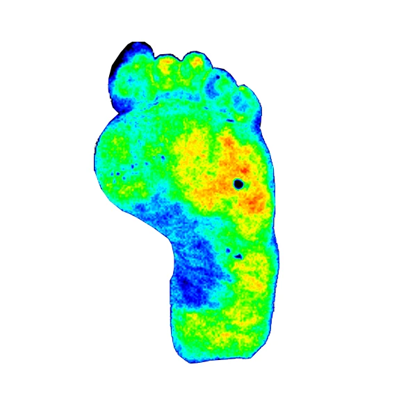
PeriCam PSI NR
Our non-contact, noninvasive imaging system uses laser speckle contrast imaging (LSCI) to deliver high-resolution tissue-perfusion images in real-time.
Without the need for contrast or tracer agents, PeriCam PSI helps you assess wound healing potential and monitor progress over time without disrupting the wound site.
Our DICOM option connects your PeriCam PSI NR to existing patient management systems for rapid and error-free patient data retrieval and instant report sharing.
Contact us
Get in touch
If you’d like to know more about how our solutions can support you in wound healing, fill out the form and we will be in touch.
LASCA and LSCI
Laser speckle contrast imaging (LSCI) is the innovative technology behind PeriCam PSI, offering non-contact, high-resolution perfusion imaging. The technique was first described by J.D. Briers and S. Webster in 1996 and referred to as laser speckle contrast analysis (LASCA). In later years, the term LSCI gained in popularity and is nowadays the more common term. However, LSCI and LASCA both refer to the same technique, providing precise and reliable perfusion data without the need for contrast agents.