Iontophoresis
Iontophoresis is a noninvasive method for administering drug ions across biological barriers, such as skin, using a low electrical current.
It is an invaluable tool for clinical researchers, particularly in the study of vascular diseases, microcirculation, and skin conditions — as well as in the development of novel therapies in these areas.
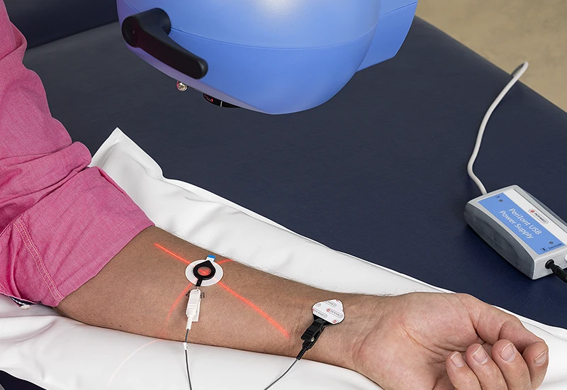
When combined with technologies like laser Doppler perfusion monitoring (LDPM) or laser speckle contrast imaging (LSCI), iontophoresis provides detailed insights into how the microcirculation reacts to drugs.
We offer iontophoresis solutions intended for research, which are compatible with PeriFlux 6000 LPDM and PeriCam PSI imagers.
How does it work?
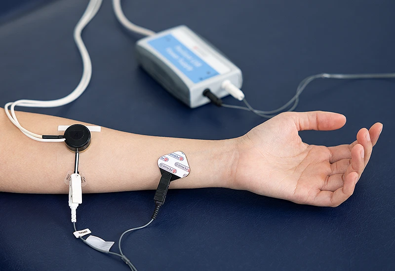
An electrode with a charge matching that of the ionized drug is placed on the skin, and another electrode with the opposite charge is positioned nearby, creating an electric field that propels the drug ions into the tissue. You can use PeriFlux 6000 LPDM or PeriCam PSI to measure the effect.
The choice of instrument depends on the needs of your clinical study. For clinical research emphasizing spatial variability or needing a broad overview of microvascular function, imaging solutions like the PeriCam PSI are probably best. For studies that require dynamic data at a single point, probe-based solutions like PeriFlux 6000 LDPM would be suitable.
Why use iontophoresis?
As the technique allows for precise and controlled delivery of ionic drugs, iontophoresis is suitable for research into the localized effect of a compound. The effectiveness of drug dispersion, however, depends on various factors, including the drug’s molecular size, charge, and concentration, as well as skin condition and local blood perfusion.
The lack of need for needles enhances measurement robustness. Without needles, there is no tissue trauma, and disruption to the local blood flow and microcirculation at the measurement site is minimal. Needle insertion can cause pain, inflammation, or localized swelling — interfering with the natural physiological state and measurement results.
Noninvasive approaches reduce the risk of infection and cross-contamination. Some people are more relaxed and comfortable without needles, leading to more stable physiological responses and consistent and reliable data.
Iontophoresis is especially suited for studying the effect of compounds on vascular systems, offering researchers a reliable way to investigate endothelial-dependent and independent responses. Leveraging iontophoresis and advanced diagnostic tools enables researchers to deepen their understanding of vascular diseases, paving the way for innovative treatments and interventions.
PeriIont for PeriFlux 6000

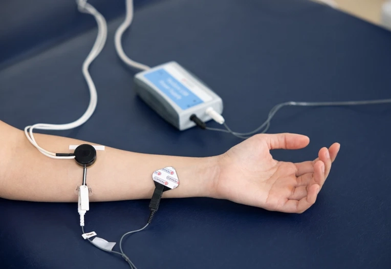
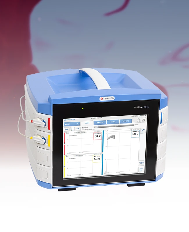
Our iontophoresis solution for PeriFlux 6000 includes everything you need, including a specialized laser Doppler probe that combines drug delivery with measurement, power supply, and Windows software to control experiments.
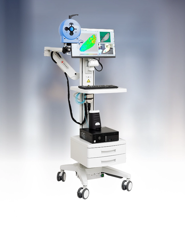
PeriIont for PeriCam PSI
Our iontophoresis solution for PeriCam PSI includes everything you need, including delivery and dispersive electrodes, power supply, and Windows software to control experiments. The positioning pillow helps to reduce noise in recordings.
Related products
Contact us
Fill out the form if you you would like to know more about iontophoresis and how it might fit into your clinical research.
LASCA and LSCI
Laser speckle contrast imaging (LSCI) is the innovative technology behind PeriCam PSI, offering non-contact, high-resolution perfusion imaging. The technique was first described by J.D. Briers and S. Webster in 1996 and referred to as laser speckle contrast analysis (LASCA). In later years, the term LSCI gained in popularity and is nowadays the more common term. However, LSCI and LASCA both refer to the same technique, providing precise and reliable perfusion data without the need for contrast agents.