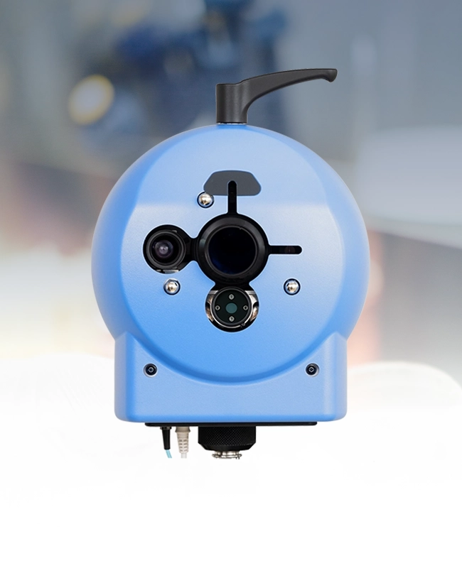
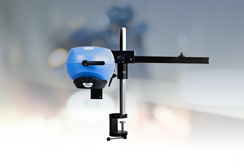
PeriCam imaging systems
Real-time perfusion imaging
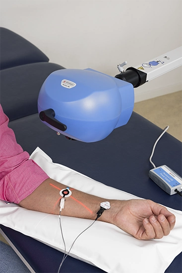
Designed for use in clinical settings, clinical research, and preclinical research, our PeriCam imagers leverage laser speckle contrast imaging (LSCI) to deliver blood perfusion data, images, and video in real-time.
All our PeriCam instruments are non-contact, noninvasive, and non-destructive, with capabilities to deliver high-resolution images of small, highly detailed areas of interest to larger parts of the body.
Evolution of medical imaging technology
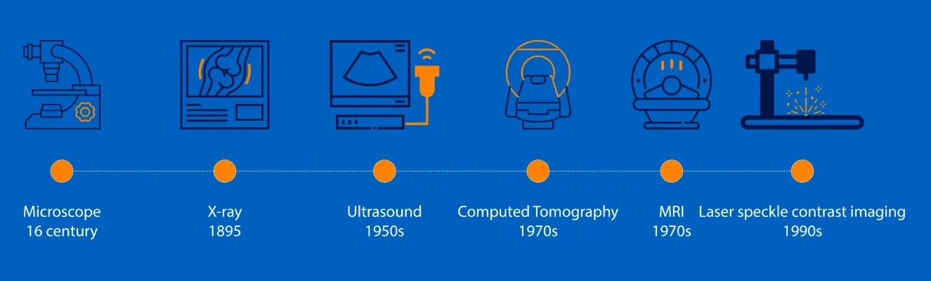
Medical imaging technology has revolutionized how we diagnose, monitor, and treat diseases. Techniques such as magnetic resonance (MRI), computerized axial tomography (CAT), and positron emission tomography (PET) leveraged emerging computing capabilities of the eighties and metabolic imaging to provide clinicians and researchers with detailed internal structures and critical insights into complex medical conditions. These technologies expanded research possibilities, enabling real-time visualization of physiological responses, disease progression, and therapeutic effects on a cellular level.
Developments in materials, AI, sensors, and computing capabilities have led to a new wave of smaller, noninvasive, non-contact instruments that support early detection, personalized treatment, and the development of innovative therapies.
In addition to size and maneuverability, one of the main advantages of modern medical imaging solutions like PeriCam is that they don’t require clinical staff to administer contrast or tracer agents. Modern imaging techniques are consequently easier to use and have less impact.
The hidden benefit, however, lies in the lack of interference with the tissue being measured. Without contrast agents or contact, tissue is measured in its natural, unaltered state. In this way, PeriCam imaging solutions reveal physiological processes as they occur, which is especially valuable for accuracy and reliability in clinical and research settings.
Data and images
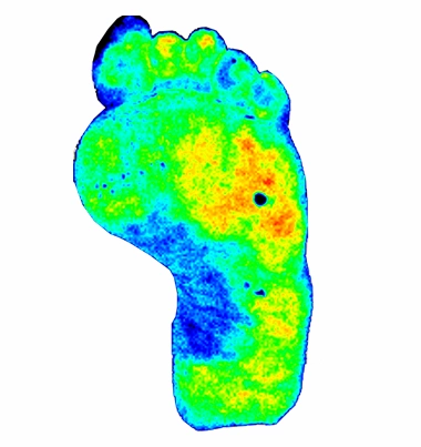
PeriCam transforms blood perfusion in actual tissue into a real-time data stream of perfusion units and interprets that data into easy-to-understand color-coded images.
In this perfusion image of a person’s foot with an ulcer, the color coding indicates lower perfusion in the blue areas compared with the green and yellow ones. Perfusion around the ulcer — red and orange areas — is elevated, indicating an inflammatory response and no severe ischemia.
Clinical application
PeriCam provides clinicians with immediate insights about blood perfusion and tissue health, which is useful for assessing wounds, evaluating microvascular function, and identifying compromised blood flow in patients with diabetes or vascular diseases. Its non-contact design and real-time visualization support clinical workflows by minimizing patient discomfort and allowing for seamless integration into regular diagnostic practices.

Clinical research
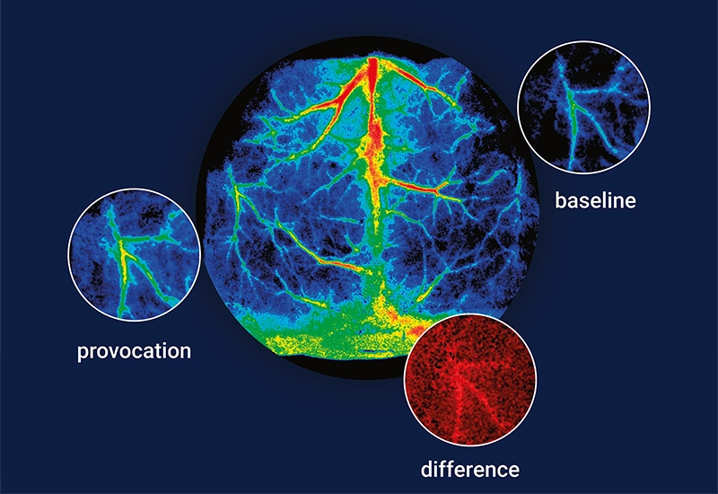
PeriCam’s precision becomes a powerful tool for studying tissue and physiological responses to therapies and provocations without the need for contrast agents or physical contact that can disrupt delicate tissues. Researchers benefit from the ability to measure tissue in its natural state.
Preclinical
PeriCam’s sensitivity to minute blood flow changes provides insights into how new compounds or interventions affect microcirculation, aiding drug development and efficacy testing.
PeriCam PSI models
To meet the highly diverse needs of researchers and clinicians, we’ve designed two PeriCam PSI models: PeriCam PSI NR and PeriCam PSI HR, both with optional zoom functionality.
Contact US
Get in touch
Fill out the form if you would like to know more about PeriCam imaging systems. We will put a local representative in touch with you shortly.
LASCA and LSCI
Laser speckle contrast imaging (LSCI) is the innovative technology behind PeriCam PSI, offering non-contact, high-resolution perfusion imaging. The technique was first described by J.D. Briers and S. Webster in 1996 and referred to as laser speckle contrast analysis (LASCA). In later years, the term LSCI gained in popularity and is nowadays the more common term. However, LSCI and LASCA both refer to the same technique, providing precise and reliable perfusion data without the need for contrast agents.