PeriFlux 6000 TcpO2 System
Measuring microcirculation with ease
TcpO2 is a noninvasive method for measuring how much oxygen the microcirculation supplies to local tissue. PeriFlux 6000 TcpO2 System generates robust and objective TcpO2 measurements, enabling you to follow patients and research subjects throughout treatment and trials, and assess the effectiveness of every intervention.
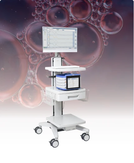
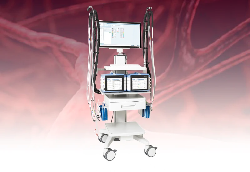
Mounted on a cart for convenient placement, transportation, and maneuverability, PeriFlux 6000 TcpO2 System supports up to eight simultaneous TcpO2 measurements.
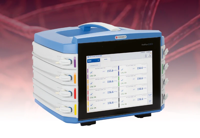
Multiple sensors and intuitive interface
Multiple sensors enable you to simultaneously measure tissue health at several points on the body— minimizing the time you spend measuring the patient.
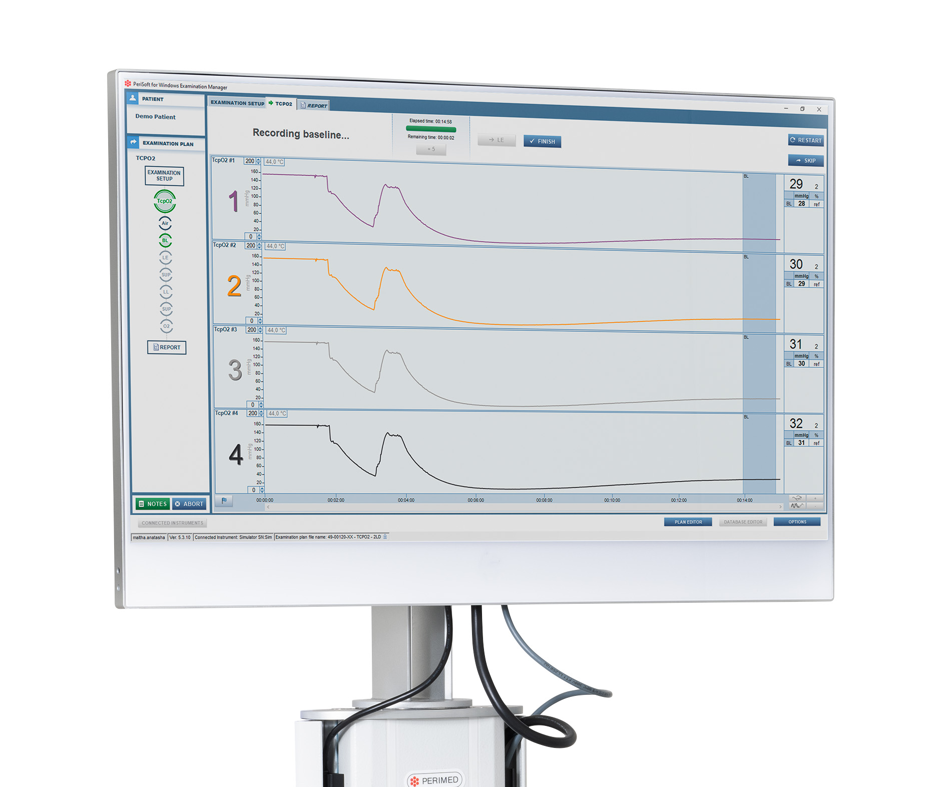
The system aids the interpretation of TcpO2 measurements through simple visualization of recorded values in real-time.
- Configured to suit your clinical/research processes.
- Interface guides you through the measurement steps.
- Each TcpO2 electrode is color-coded to match the user interface, supporting ease of use and identification of microcirculation issues.
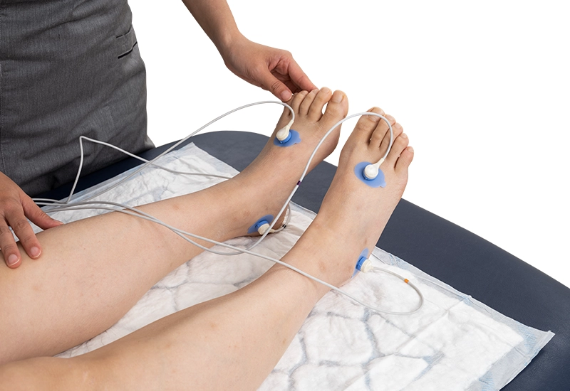
Noninvasive sensors
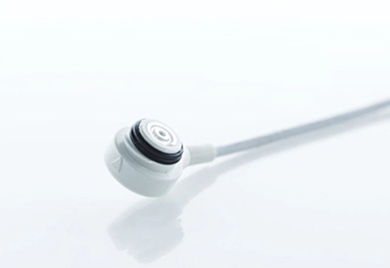
Owing to their sensitivity, our system uses Clark-type electrodes for conducting TcpO2 measurements.
These sensors are noninvasive and are placed on the body with self-adhesive (single-use) fixation rings. The electrode heats the measurement site to encourage any available oxygen in the microcirculation to diffuse through the skin.

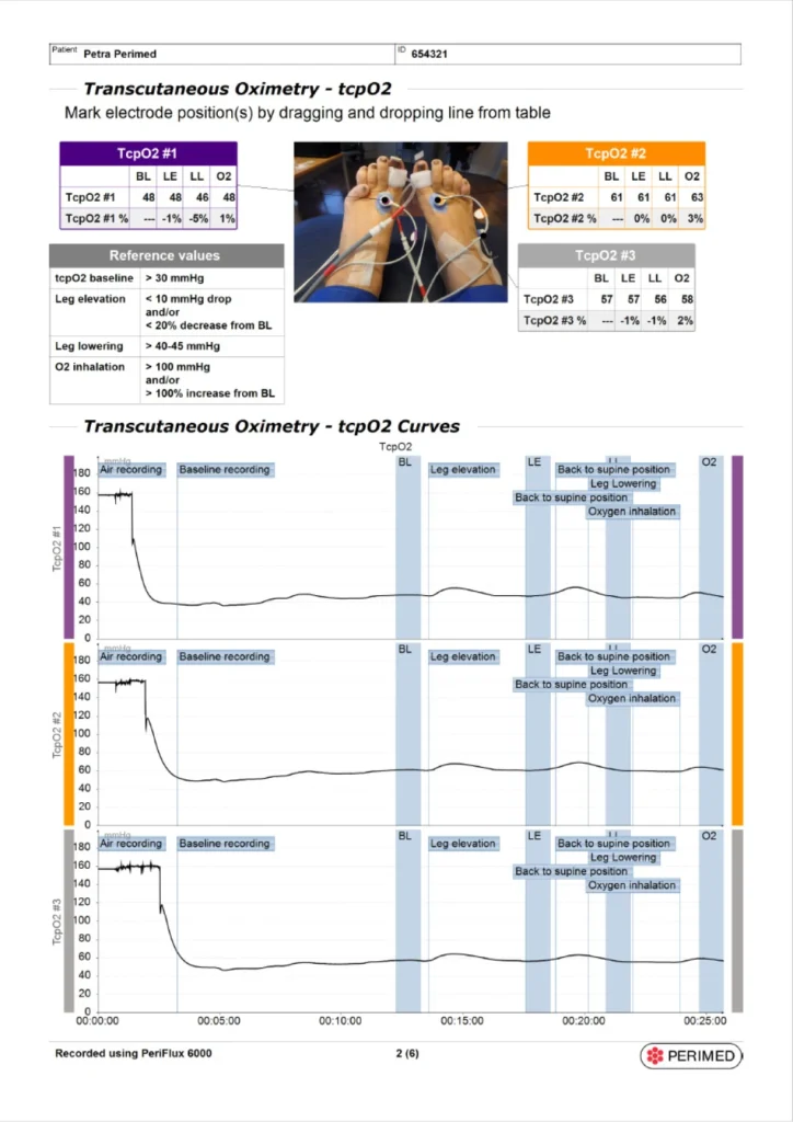
The report
All measurements — and any images you take for documentation purposes — are conveniently packaged in a comprehensive report, which can be securely transferred to other systems using DICOM. For research purposes, it is possible to export the report to XML.
TcpO2 applications
In addition to being a recommended method [1] for quantifying the severity of ischemia, determining critical limb-threatening ischemia (CLTI) prognosis, assessing eligibility for hyperbaric oxygen treatment (HBOT), and the effectiveness of HBOT and revascularization, TcpO2 delivers useful metrics that aid wound healing prediction, amputation level decision making, and peripheral artery disease (PAD) diagnosis for people with calcified arteries or amputated toes.

TcpO2 applications

Severity of ischemia

Wound healing prediction

HBOT eligibility

HBOT effectiveness

Revascularization effectiveness

Amputation level determination
PeriFlux 6000 TcpO2 — components
Mounted on a cart for convenient placement, transportation, and maneuverability, PeriFlux 6000 TcpO2 System supports up to eight simultaneous TcpO2 measurements.
- Guided workflow application software — PSW ExM.
- TcpO2 unit — up to eight measurement channels for comparable measurements.
- Clark electrodes — standard for TcpO2 measurements.
- All-in-one computer — runs the application software and provides the user interface.
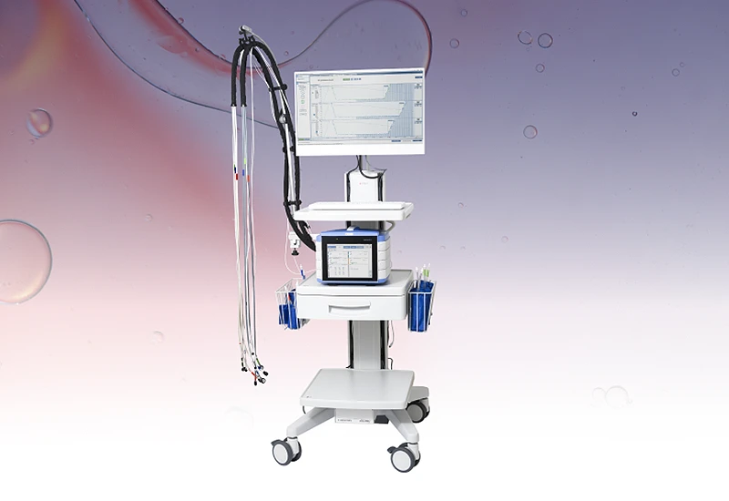
- Can be upgraded to PeriFlux 6000 Combined to expand capabilities to include pressure measurements such as toe pressure, ankle pressure, and skin perfusion pressure (SPP).
- Delivers all measurements, and images taken for documentation purposes, in an automatically generated patient report.
- Capability to export to XML for research purposes.
- Color-coded electrodes and interface to ensure ease of use.
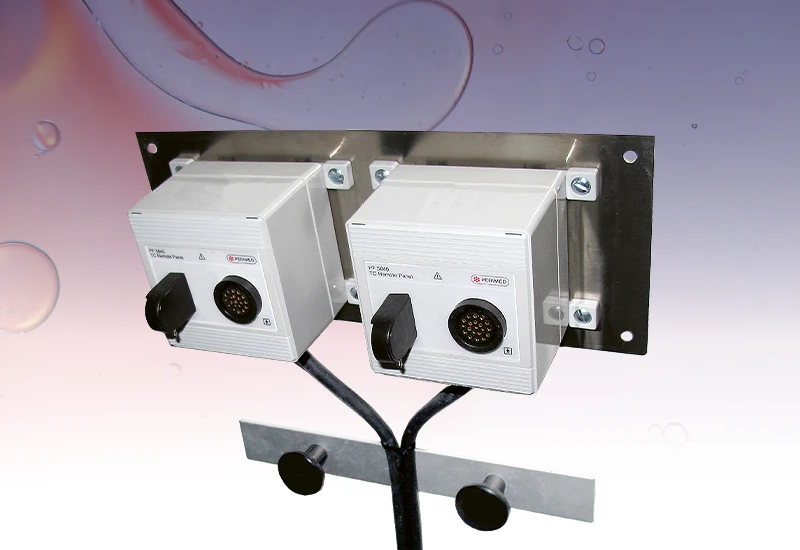
Options
- Remote panels for use in multi-place hyperbaric oxygen chambers.
- DICOM/HL7 connectivity — securely transfer data to other systems.
Related products
We designed PeriFlux 6000 TcpO2 to provide clinicians and researchers with robust TcpO2 microcirculation measurements. In addition to our TcpO2 solution, we offer PeriFlux 6000 in several additional configurations: a combined TcpO2 and pressure system, a pressure-only system, and a compact TcpO2 standalone.
Contact us
Get in touch
Get in touch if you would like to know more about PeriFlux 6000 TcpO2 System and how it might fit into your clinical processes.
References
- Conte MS, Bradbury AW, Kolh P, et al. Global Vascular Guidelines on the Management of Chronic Limb-Threatening Ischemia. European Journal of Vascular and Endovascular Surgery. 2019;58(1):S1-S109.e33. doi:10.1016/j.ejvs.2019.05.006