Cerebral blood flow monitoring
Cerebral blood flow monitoring leverages blood perfusion to understand and assess brain function. It is a popular biomarker among neuroscientists as it is indicative of many neurological conditions — such as stroke, traumatic brain injury (TBI), and Alzheimer’s disease. With proper measurement tools, cerebral blood flow monitoring is suitable for assessing disease progression, the impact of therapeutic interventions, and the brain’s ability to recover.

Overcoming data collection challenges
Small or subtle variations in blood flow, especially in microcirculatory networks, are often critical for studying conditions like stroke, traumatic brain injury, and neurodegenerative diseases. These minute changes can be hard to measure without advanced tools. Leveraging laser speckle contrast imaging (LSCI), PeriCam PSI aims to overcome this challenge by delivering high-resolution images up to 10 μm/pixel, providing researchers with an effective means to capture minute variations in blood flow.
Continuous real-time data is a prerequisite for tracking dynamic processes like neurovascular coupling or the sudden influx of blood flow following ischemia that occurs in stroke, heart attack, and revascularization surgery. PeriCam PSI records these processes at a frequency of up to 100 images per second, capturing events at the required level of detail — spatially and temporally.
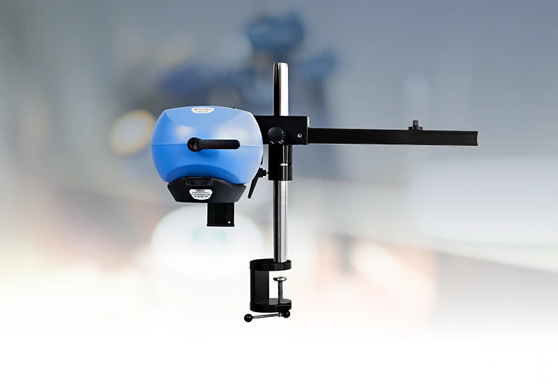
Physiological factors like breathing, movement, or pulsation and external factors, such as vibration or ambient light fluctuation, can introduce artifacts into the data if they are not accounted for. PeriCam PSI is delivered on a high-quality arm that helps to prevent movement artifacts and automated background compensation ensures robust measurements under varying lighting conditions.
Traditional imaging techniques often require contact with the tissue or contrast agents, which can disrupt natural physiology and compromise data integrity. PeriCam PSI helps address this challenge by providing clear insights into blood flow dynamics without physically affecting the tissue.
PeriCam PSI in practice
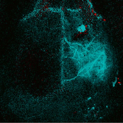
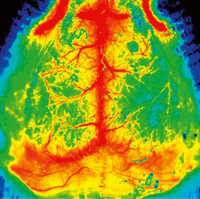
PeriCam PSI’s high spatial resolution enables researchers to visualize an entire mouse brain, making it possible to characterize and precisely locate ischemic injury.
Mouse brain. PeriCam PSI HR Courtesy Dr Offenhauser, Charité, Berlin, Germany.
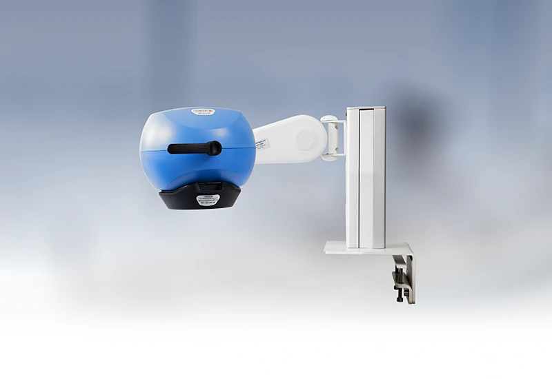
PeriCam PSI HR

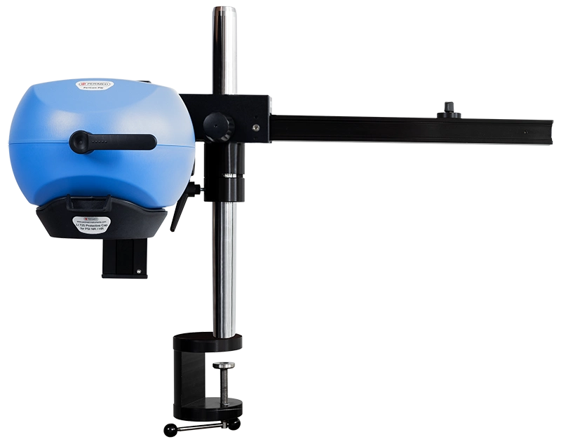
PeriCam PSI leverages laser speckle contrast imaging (LSCI) to assess blood perfusion in tissue at the microcirculation level with detail and precision. PeriCam PSI HR (high resolution) works from a fixed distance and is specifically equipped to measure small animals such as rats and mice.
- Rapid data analysis, delivering perfusion graphs, images, and video recordings in real-time.
- Regions of interest (ROIs) and time periods of interest (TOIs) that can be defined and updated before, during, and after recording.
- Frequency of up to ~100 images per second.
- Automated background compensation for robust measurements under varying lighting conditions.
- Suitable for longitudinal studies.
- High-quality arm with good maneuverability to ease positioning, and stability to prevent movement artifacts.
- Wide-angled color camera for documentation of the measurement area and its surroundings, easing post-measurement analysis.
- Non-contact, non-destructive, and noninvasive
Related products
Contact us
Get in touch
Our history is deep-rooted in research and we are here to support you with the tools and expertise needed for effective data collection in preclinical research. Contact us to learn more about how our solutions can support you and start changing outcomes.
LASCA and LSCI
Laser speckle contrast imaging (LSCI) is the innovative technology behind PeriCam PSI, offering non-contact, high-resolution perfusion imaging. This technique, sometimes referred to as laser speckle contrast analysis (LASCA), provides precise and reliable data without the need for contrast agents.