Middle cerebral artery occlusion
Middle cerebral artery occlusion (MCAO) is a widely used preclinical model for studying ischemic stroke. By replicating the effects of reduced blood flow to brain tissue, it induces localized ischemia and potential tissue damage.
he MCAO model is instrumental in stroke research, understanding the physiological responses and testing the efficacy of new therapeutic approaches before they reach clinical trials. MCAO studies require precise data collection to capture the subtle, time-sensitive changes in blood flow and tissue perfusion that occur immediately after occlusion and during recovery.
Overcoming challenges
The degree of ischemia can vary widely depending on the method used to induce occlusion. Achieving reproducibility in the infarct’s size and location is also difficult, and small variations can alter the outcome.
MCAO research relies on precise timing to capture changes during the critical phases of stroke onset, progression, and recovery.
Monitoring microcirculatory changes in real-time without introducing artifacts is also complex. Traditional imaging techniques often require contact with the tissue or contrast agents, which can disrupt natural physiology and compromise data integrity. PeriCam PSI helps address this challenge by providing clear insights into blood flow dynamics without physically affecting the tissue.
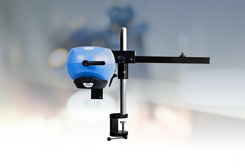
PeriCam PSI HR

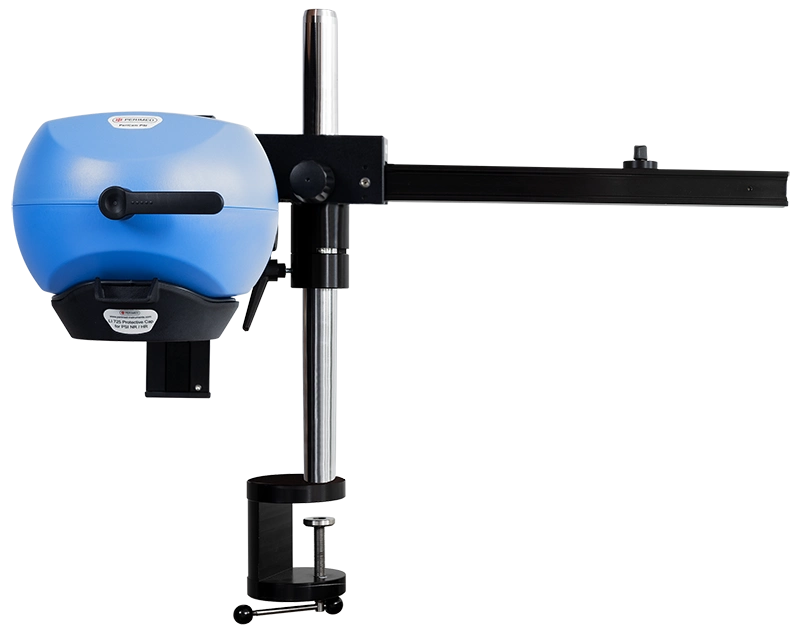
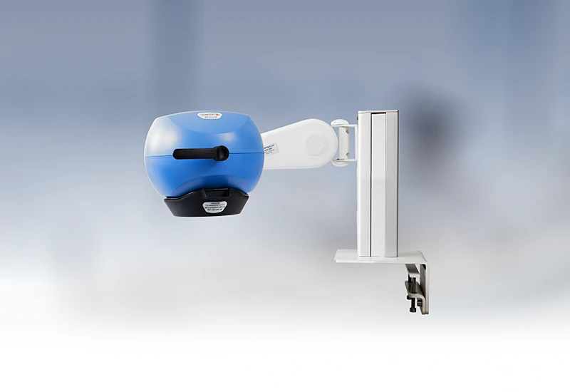
PeriCam PSI leverages laser speckle contrast imaging (LSCI) to assess blood perfusion in tissue at the microcirculation level with detail and precision. PeriCam PSI HR (high resolution) works from a fixed distance and is specifically equipped to measure small animals such as rats and mice.
- Rapid data analysis, delivering perfusion graphs, images, and video recordings in real-time.
- Regions of interest (ROIs) and time periods of interest (TOIs) that can be defined and updated before, during, and after recording.
- Frequency of up to ~100 images per second.
- Automated background compensation for robust measurements under varying lighting conditions.
- Suitable for longitudinal studies.
- High-quality arm with good maneuverability to ease positioning, and stability to prevent movement artifacts.
- Wide-angled color camera for documentation of the measurement area and its surroundings, easing post-measurement analysis.
- Non-contact, non-destructive, and noninvasive.
PeriCam PSI in practice
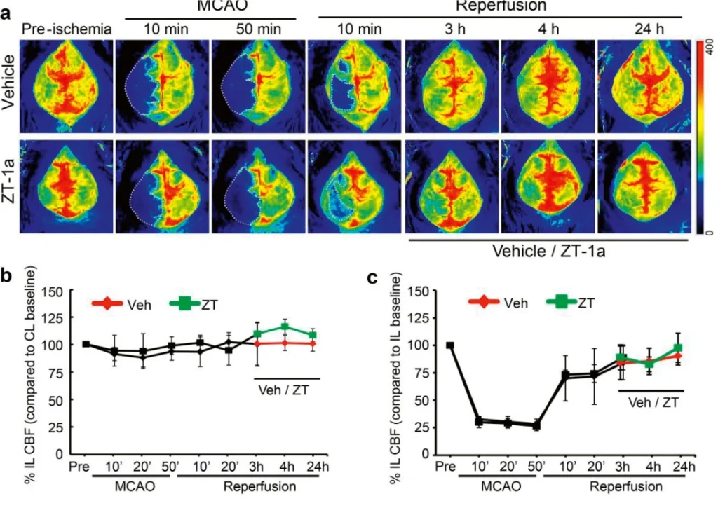
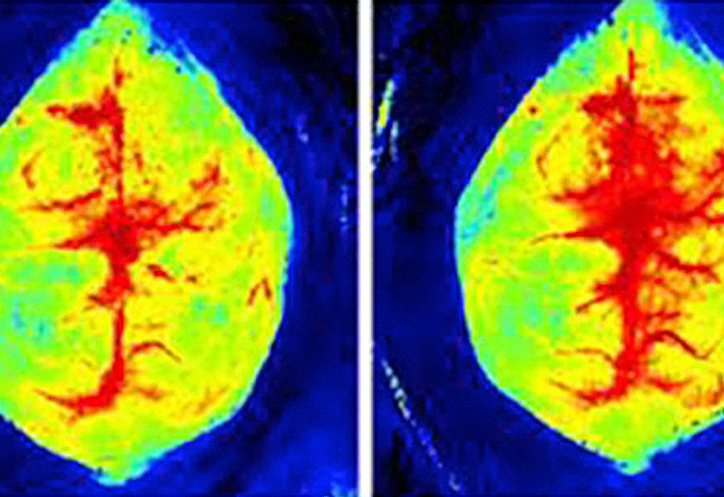
Perfusion images generated by PeriCam PSI show a mouse brain before, during, and after stroke induction, the graphs show quantified changes in blood perfusion of the ipsilateral and contralateral sides for both the treated and untreated animal.
Related products
Contact us
Get in touch
PeriCam PSI supports stroke research by providing real time data with noninvasive, non-contact, and non-destructive methods. For more information, fill out the form and we will be in touch with you shortly.
LASCA and LSCI
Laser speckle contrast imaging (LSCI) is the innovative technology behind PeriCam PSI, offering non-contact, high-resolution perfusion imaging. The technique was first described by J.D. Briers and S. Webster in 1996 and referred to as laser speckle contrast analysis (LASCA). In later years, the term LSCI gained in popularity and is nowadays the more common term. However, LSCI and LASCA both refer to the same technique, providing precise and reliable perfusion data without the need for contrast agents.