Pulse volume recording — PVR
Pulse volume recording (PVR) is a noninvasive method for evaluating blood flow in peripheral arteries based on air plethysmography. PVR is suitable for localizing segmental disease in patients with non-compressible arteries.
For every heartbeat, a subtle expansion and contraction occurs in the arteries, causing limb volume to vary slightly. PVR is a measurement of the volume change.

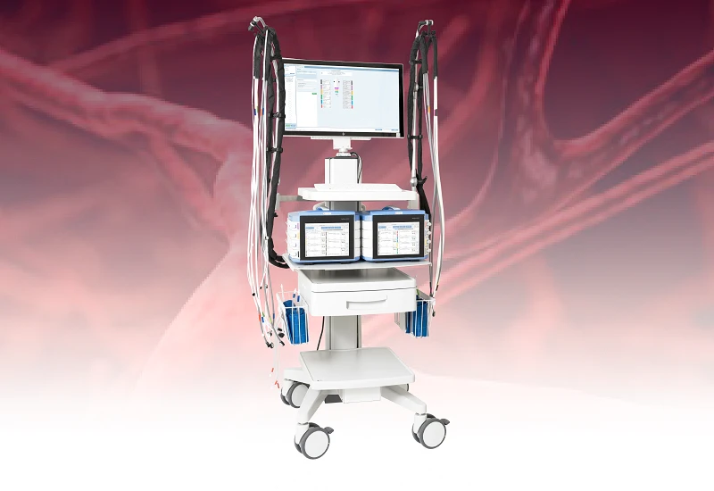
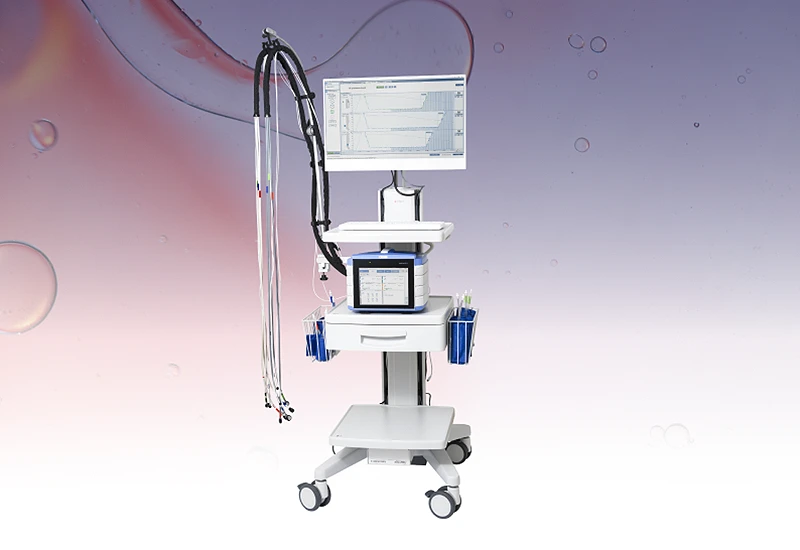
PVR with PeriFlux 6000
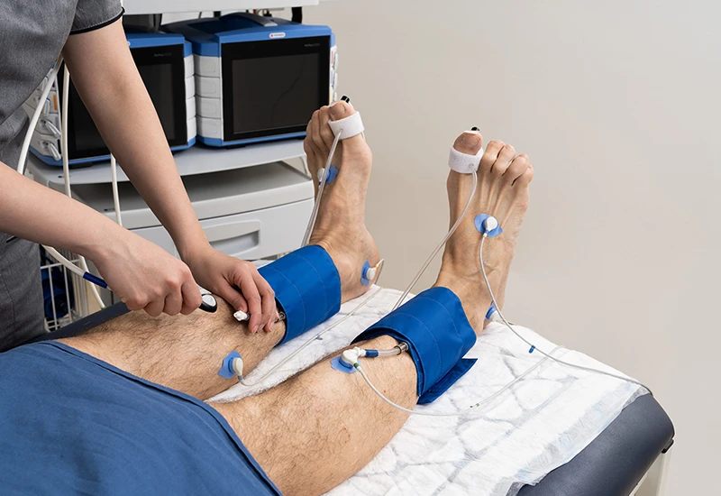
PVR relies on air plethysmography, a technology that measures volume changes in the limb using pressure cuffs. The pressure cuff is wound around the limb being measured, such as the thigh, calf, or ankle. The cuff is partially inflated to 60 mmHg to avoid compressing the artery while remaining sensitive to volume changes.
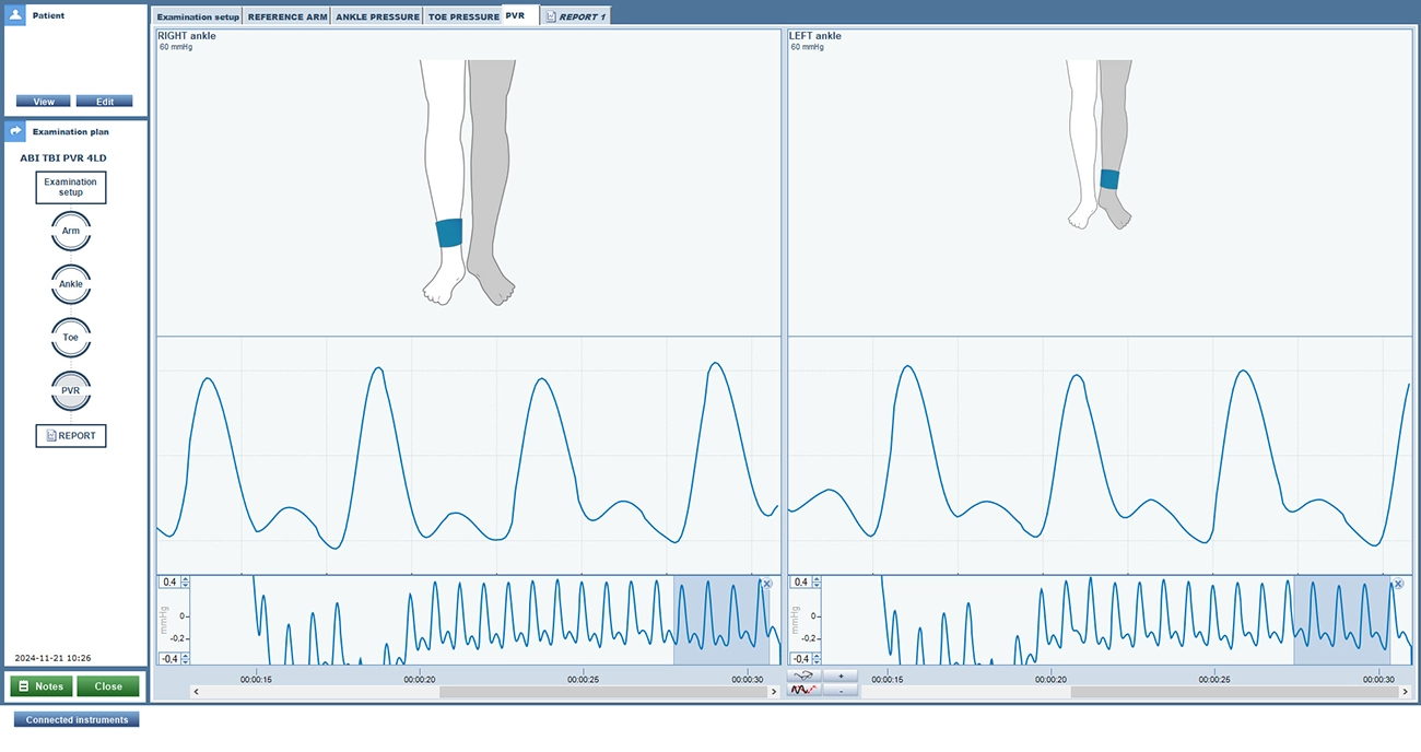
With each cardiac cycle, blood pulses through the arteries, causing the limb to expand slightly. The cuff senses these minute changes in limb volume. PeriFlux 6000 leverages advanced signal processing to generate a pulse volume waveform based on the volume change.
Interpreting PVR
The pulse volume waveform offers information about the condition of the arteries. A healthy artery produces a sharp, well-defined waveform. Lack of a dicrotic notch or a dampened waveform may indicate reduced blood flow or arterial stiffness.
Clinical advantage
PVR is noninvasive, reproducible, and can assess blood flow at multiple levels of the limb in a single session. The test is easy to perform, quick, and does not require contrast agents.
PVR is often used when ankle pressure is unreliable due to non-compressible arteries and combined with other diagnostic methods, such as toe pressure and other microcirculation measurements such as TcpO2 or skin perfusion pressure (SPP) — for a comprehensive vascular assessment.
Related products and applications
Contact us
Get in touch
If you would like to know more about how to carry out pulse volume recording (PVR), its application, or our related products, fill out the form and we will be in touch with you shortly.