


The power of combining laser Doppler with TcpO2 — one protocol, multiple insights

by Sara Gyllensporre (Clinical Application Specialist)
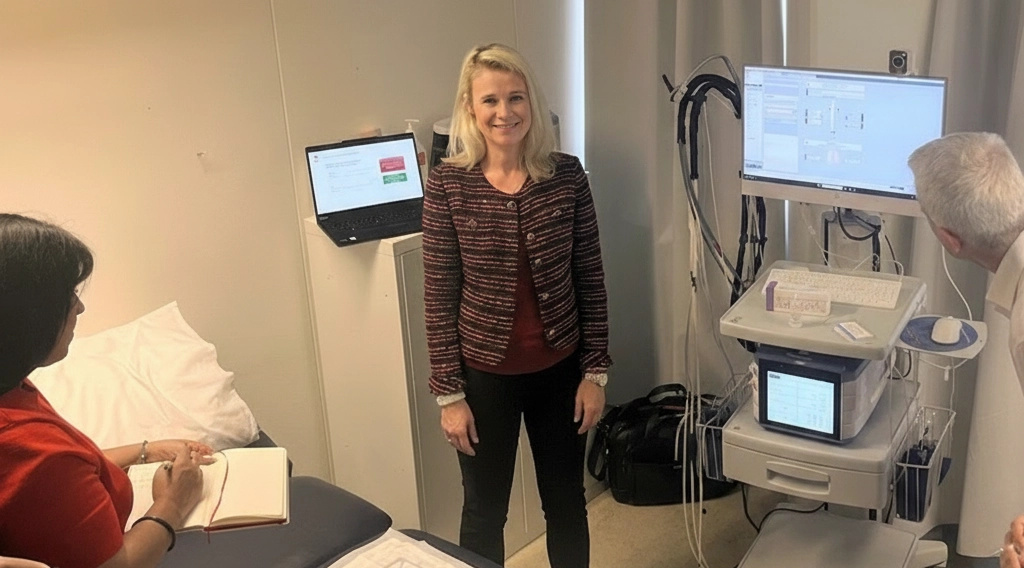
In a recent training session with our distributors, I demonstrated a simple yet powerful protocol that combines baseline and provocation measurements for Laser Doppler (LD) perfusion and transcutaneous oxygen (TcpO2).
LD perfusion measurements are straightforward as they don’t require occlusion; just place the probe and measure. Another aspect is time. Because LD and TcpO2 do not interfere with each other, you can record baselines simultaneously.
The real power, however, comes from the combination of LD, LD with heat provocation, TcpO2, TcpO2 oxygen challenge, and additional TcpO2 provocations to deliver multiple complementary insights into a person’s vascular status.
In this post, I take a closer look at the LD + TcpO2 protocol by briefly explaining the underlying physiology and presenting a couple of example patient cases that show how the individual measurements compare with the combined approach. If you’re interested in using this protocol with PeriFlux 6000, please get in touch by filling out the form at the end of this post.
Laser Doppler in macro- and microcirculation measurements
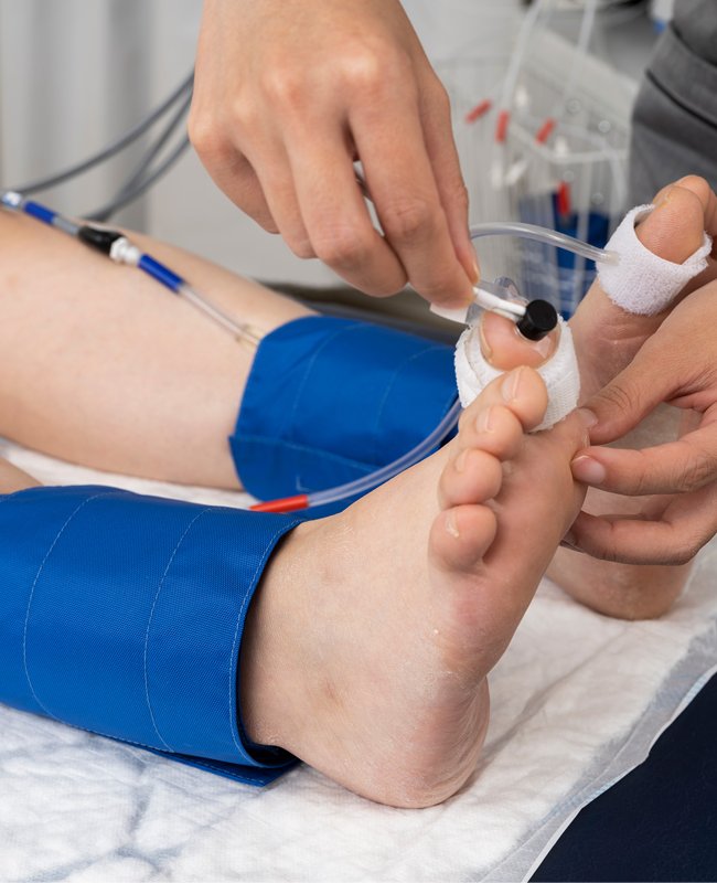
In traditional macrocirculation measurements, like ABI and TBI, the laser Doppler probe is usually placed on the toe — distal to the limb. During cuff inflation, occlusion, and release, the probe measures underlying blood flow, and advanced signal processing identifies when occlusion occurs and when blood flow returns.
Laser Doppler probes can also be used directly without occlusion to assess local cutaneous microvascular function. In isolation, LD perfusion provides some insight, but when combined with provocations and other microvascular protocols such as TcpO2, its value increases significantly.
The underlying physiology
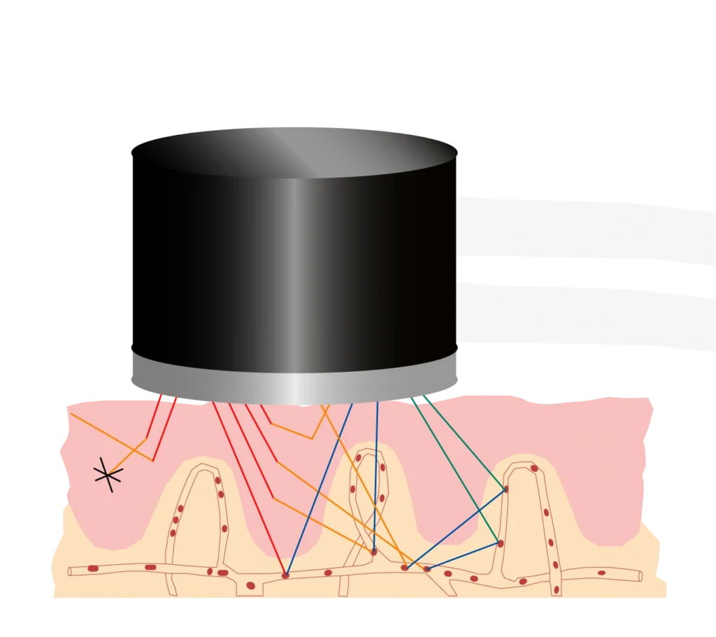
A laser Doppler probe placed directly on the skin emits low-power laser light into the underlying tissue. Some light is absorbed, and the rest is scattered. When scattered light encounters a stationary structure, it changes direction (illustrated by the orange lines). When scattered light encounters a moving red blood cell (RBC), it changes frequency — due to the Doppler effect — as well as direction (illustrated by the blue and green lines). Perfusion is calculated based on the frequency spectrum of the light returning to the probe.
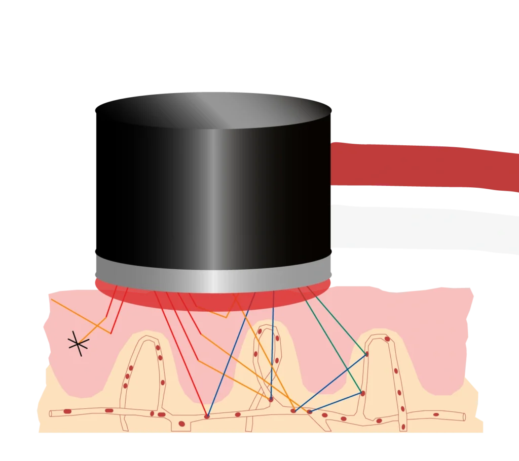
When you heat the skin to, for example, 44°C, the small blood vessels in the tissue dilate, increasing cutaneous microvascular blood flow. In a healthy person, the response is usually immediate, and perfusion typically rises rapidly. In people with impaired microvascular function, this response may be delayed, attenuated, or otherwise impaired.
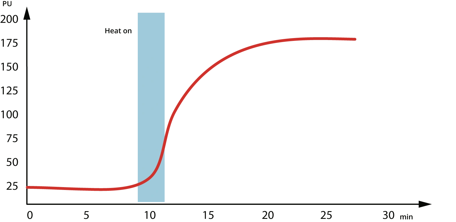
Interpreting curves — Patient A
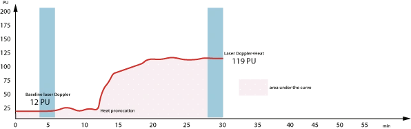
Patient A responded clearly to heat. Perfusion increased almost immediately after probe heating and plateaued about 10 minutes later.
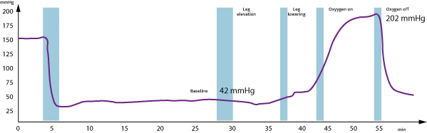
At 42 mmHg, the patient’s baseline was borderline*, suggesting inflammation, edema, or possibly both. However, the patient’s response to an oxygen challenge showed a clinically significant increase in TcpO2 [1], which, together with the strong vasodilatory response observed in the laser Doppler heat measurement, suggests preserved wound-healing potential.
*The Global Vascular Guidelines on Chronic Limb-Threatening Ischemia [2] indicate TcpO2 <40 mmHg is associated with increased wound complications. TcpO2 values above this threshold are generally associated with favorable tissue oxygenation.
Typical thresholds for LD with heat
Clinically established LD thresholds comparable to those used for ABI, skin perfusion pressure (SPP), and TcpO2 do not exist. Several factors contribute to this: underlying physiological variation, different measurement technologies, and diverse methods for signal interpretation.
In my experience, typical baseline values measured with a PeriFlux 6000 instrument range from 5-50 PU and rise to 100-300 PU during heat provocation. I tend to look at the area under the curve, the percentage increase from baseline to plateau, response time, and the time to peak or plateau, as these reflect microvascular reactivity and vasodilatory capacity.
Interpreting curves — Patient B
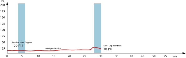
Patient B had a non-healing wound. The clear lack of response to heat indicates impairment. In such cases, I first validate the measurement by checking that the probe is properly fixed to the skin. Sometimes, I follow up with a second measurement at a different site — one that hasn’t been exposed to heat (non-dilated).
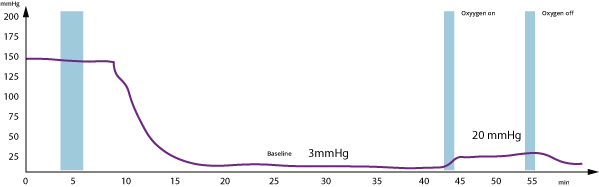
Patient B’s TcpO2 baseline was below the generally recommended wound-healing threshold of 40 mmHg, and oxygen response was minimal.
Together, these insights indicate impaired microvascular responsiveness.
Interpreting curves — Patient C
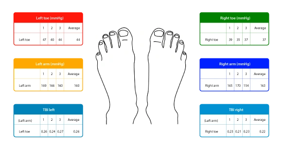
Patient C is a 90-year-old woman with diabetes, foot discoloration, and low ABI. We first measured toe pressure and TBI.
Taking an average of three toe pressure measurements, at 0.26 on the left and 0.22 on the right, TBI is low.
At these levels, the findings are suggestive of PAD.**
** In the list of recommendations for PAD diagnosis, the IWGDF Guidelines on the Prevention and Management of Diabetic Foot Disease 2023 [3] states: PAD is less likely in the presence of ABI 0.9-1.3; TBI ≥ 0.70.
To determine the best course of therapy, we followed up with TcpO2, laser Doppler baseline, and a laser Doppler heat provocation. In this case, the patient had no wounds, so we placed the LD probes and TcpO2 electrodes as shown in the illustration below. If a wound is present, I recommend placing these sensors as close to the wound as possible.

One of the most important things to remember when carrying out multiple measurements is the probe temperature. A toe pressure measurement heats the probe, so turn off the heat and let the probe cool before performing the baseline laser Doppler measurement.
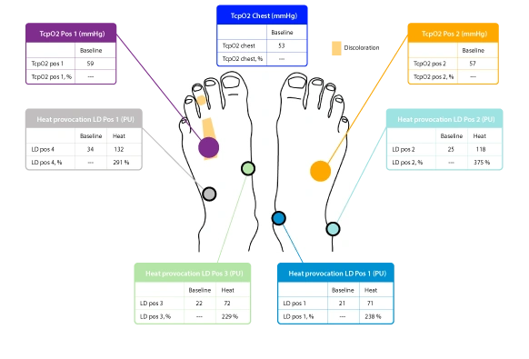
Although toe pressure was markedly reduced, baseline limb TcpO2 was above 50 mmHg and comparable to chest-level TcpO2, indicating preserved oxygen delivery. This apparent discrepancy may reflect reduced arterial pressure partly offset by collateral circulation and local blood-flow regulation, allowing the microcirculation to maintain adequate tissue oxygenation.
In addition to the low macrovascular indices, the patient had skin discoloration on the left foot. Visually and structurally, the limb looked compromised.
Application in clinical research
The combined approach of TcpO2, laser Doppler, and heat provocation may be particularly valuable in studies of peripheral artery disease (PAD), diabetes, wound healing, reconstructive surgery, and other conditions where both oxygen delivery and microvascular responsiveness play important roles. By capturing synchronized physiological data, researchers can investigate whether perfusion changes are accompanied by corresponding changes in tissue oxygenation and explore potential biomarkers of tissue viability and healing potential.
From a practical perspective, simultaneous measurements also simplify study workflows, reduce examination time, and minimize patient burden.
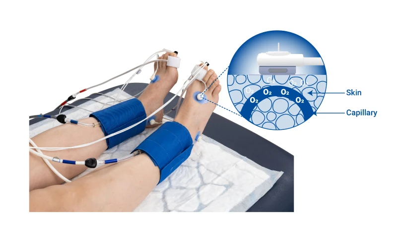
PeriFlux 6000
LD+Heat/TcpO2 is compatible with our PeriFlux 6000 Combined (laser Doppler and TcpO2) configurations. If you are interested in obtaining a protocol for your instrument, please let me know by filling out the form below.
References
[1]. Fife, C. E., Smart, D. R., Sheffield, P. J., Hopf, H. W., Hawkins, G., & Clarke, D. (2009). Transcutaneous oximetry in clinical practice: consensus statements from an expert panel based on evidence. Undersea & hyperbaric medicine : journal of the Undersea and Hyperbaric Medical Society, Inc, 36(1), 43–53.
[2]. Conte, M. S., Bradbury, A. W., Kolh, P., White, J. V., Dick, F., Fitridge, R., Mills, J. L., Ricco, J. B., Suresh, K. R., Murad, M. H., & GVG Writing Group (2019). Global vascular guidelines on the management of chronic limb-threatening ischemia. Journal of Vascular Surgery, 69(6S), 3S–125S.e40. https://doi.org/10.1016/j.jvs.2019.02.016
[3]. IWGDF Guidelines on the Prevention and Management of Diabetic Foot Disease 2023; lntersocietal 2023 PAD Guidelines
https://iwgdfguidelines.org/wp-contenUuploads/2023/07/IWGDF-2023-05-PAD-Guideline.pdf
Clinical Application Specialist
With over 16 years at Perimed, Sara has held multiple roles and developed a broad understanding of medtech and its application in healthcare settings. She holds a degree in pharmacology and an MSc in molecular biology and plays a vital role in ensuring technology meets the needs of clinical practice, supported by strong relationships with clinicians across Europe.
"*" indicates required fields
Disclaimer
The views expressed in this blog post are those of the author and do not necessarily represent the official position of Perimed. The content is provided for informational purposes only and is not intended to constitute clinical claims or medical advice. Clinical decisions should always be based on professional judgment and applicable clinical guidelines.