PeriCam PSI NR
Real-time perfusion imaging for clinical use
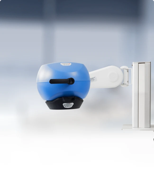
PeriCam PSI NR is a high-resolution, laser-based, noninvasive instrument that leverages laser speckle contrast imaging (LSCI) to deliver blood perfusion measurements with precision in real-time.


Designed for use in clinical and clinical research settings, PeriCam PSI NR is suitable for monitoring perfusion before, during, and after surgery, in clinical assessment, and in the development of novel drugs and therapies.
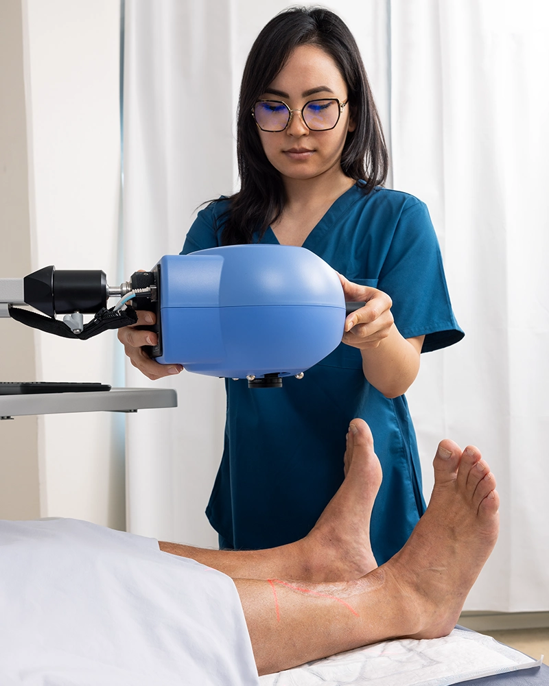
Measurement is noninvasive — no need for contrast agents — and non-contact. The instrument can be mounted on a cart or an adjustable table arm, and supports measurement of patients and study subjects from a distance of 13.0 cm up to 41.5 cm.
No contrast agents
Without the need for contrast agents, PeriCam PSI NR offers several advantages:
Patient safety — eliminates the risk of adverse reactions or allergies to agents, which can be problematic for people with sensitivities or compromised health.
Versatile application — supports repeated measurement on the same patient/study participant over short intervals. Sedation and dosage frequency of certain contrast agents can limit the number of times they can be administered.
Streamlined workflow — removes the pre-procedural steps associated with administering contrast agents, which may contribute to patient throughput and reduce data collection time.
Cost-efficiency — removes the cost of the agent itself and the resources required to administer it.
Regulatory — clinical procedures and trials that do not require contrast agents may face fewer regulatory hurdles, simplifying approval and adoption within healthcare facilities.
Application areas
PeriCam PSI NR is a versatile tool for clinical assessment and clinical research, providing high-resolution, real-time tissue perfusion critical to the understanding of disease mechanisms. Its non-contact noninvasive imaging technology supports measurement of small areas of interest as well as larger areas of a person’s body.
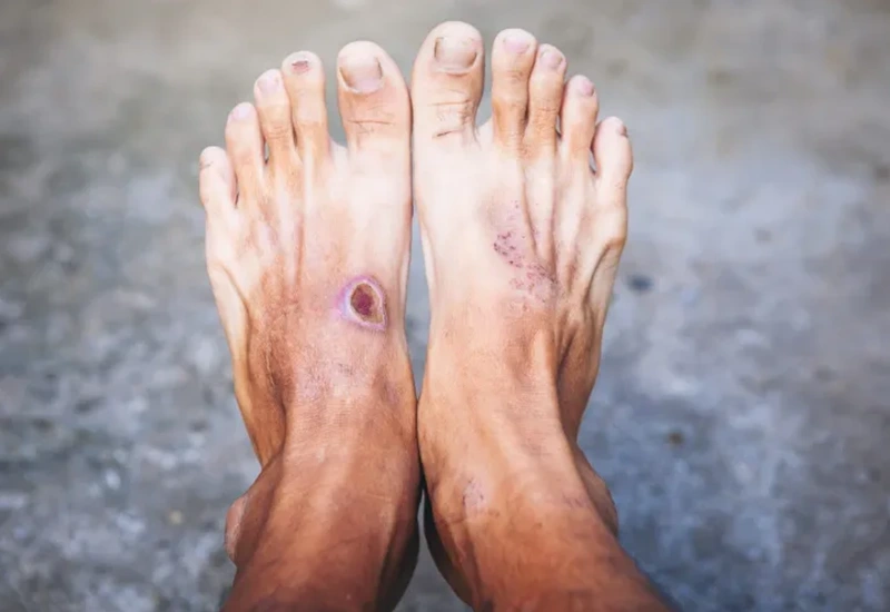
Diabetic foot
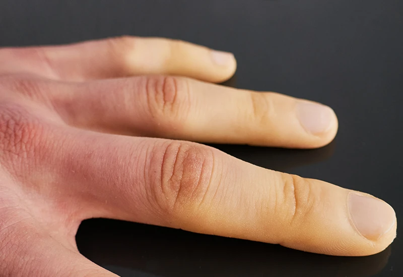
Raynaud’s Phenomenon
Cosmetics

Breast reconstruction

Vascular surgery
Drug testing
Data and images
PeriCam PSI NR converts blood perfusion in tissue into a continuous, real-time data stream of perfusion units. The included PIMSoft software translates these measurements into clear, color-coded images and graphs, providing an instant and highly visual interpretation of the underlying data.
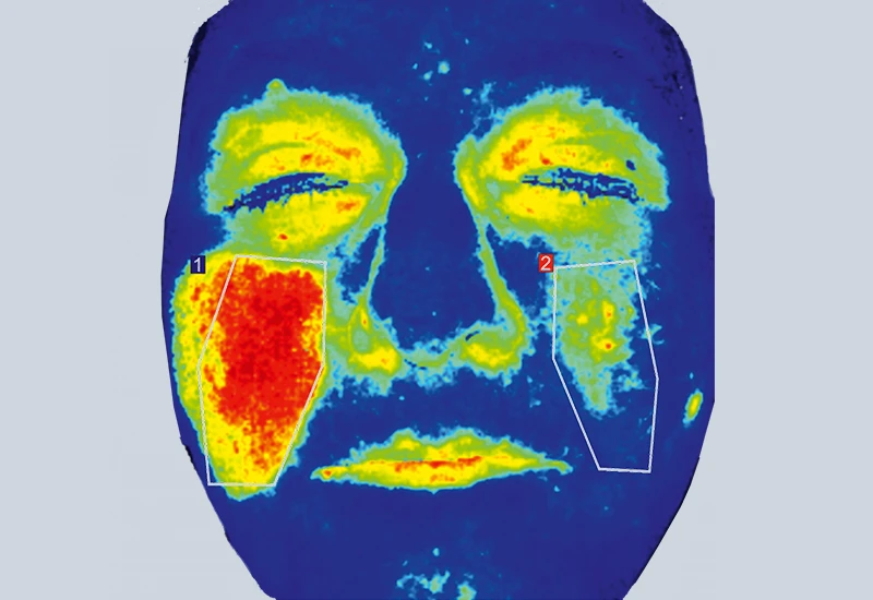
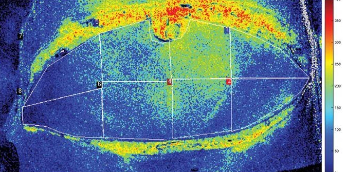
In the default color-coding system used by PeriCam PSI NR, blue/cyan indicates areas of low perfusion, with the warmer (red/orange) colors indicating areas of high perfusion.
The image, which comes from a study[1] to evaluate LSCI as a predictive technology for postoperative complication, illustrates how blood flows through a transplantation flap. The flap has been divided into regions of interest (ROIs) based on Hartrampf zones. The perforator is found in ROI I. The color coding indicates a well-perfused area above the perforator. In this case, ROIs 1-4 are viable for reconstruction purposes, while the other parts of the flap don’t have enough blood flow to be useful.
High resolution
PeriCam PSI NR supports a flexible range of measurement distances from 13 cm to 41.5 cm, with a measurement area of up to 20 x 24 cm. Blood perfusion in tissue is visualized in real-time with a pixel width of 33 µm or 10 µm with zoom functionality (the lower the pixel width, the greater the number of pixels per cm, the higher the resolution).
Easy to position
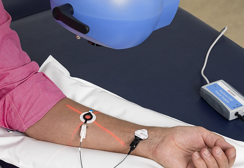
PeriCam PSI NR utilizes visible laser light to project crosshairs onto the skin of the patient/study participant at the measurement site. This feature enhances the accuracy and precision of measurements in several ways:
Laser projection — the visible laser light creates well-defined crosshairs, ensuring that the point where measurement occurs is visible and the instrument is correctly positioned to measure the desired targeted area.
Guiding placement — the crosshairs enable users to adjust the instrument’s position before taking measurements. This feature is crucial for good clinical and clinical research outcomes that require accurate assessment of tissue perfusion.
User confidence — the clear visual indicator is reassuring for clinicians and researchers, especially in dynamic environments where time is limited and accuracy crucial

Features
Regions of interest (ROIs)
PeriCam PSI NR allows you to define regions of interest (ROIs). This feature helps you to see how perfusion changes for a specific area on the patient/study participant, supporting consistency in the assessment of wound healing or treatment effectiveness, for example. ROIs can be customized in shape and size within PIMSoft.
Time periods of interest (TOIs)
In addition to regions of interest you can create time periods of interest — TOIs. This feature is valuable for documenting when events, such as drug delivery or a provocation, occur. By setting TOIs in PIMSoft, you can capture and analyze perfusion data to identify immediate and delayed physiological effects.
Continuous autofocus
This feature is designed to help you during measurement setup by automatically adjusting the camera focus to compensate for tissue movement or changes in the instrument’s positioning — ensuring every image is sharp and precisely focused on your measurement area.

Color coding
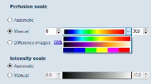
PeriCam PSI NR uses color coding to represent perfusion levels in a visually intuitive way so that the data is easy to interpret. Different schemes are built into the system. You can also define your own schemes to suit your preferences, the needs of a study, or clinical best practices.
Customizable color scheme
In addition to the default schemes, you can create your own scheme. This is a useful feature if you need to emphasize specific perfusion ranges or mimic an established color convention.
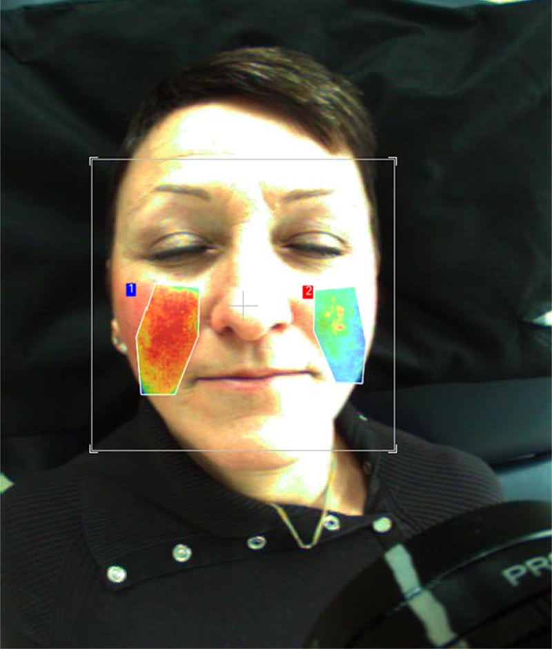
Perfusion overlay
This feature enables perfusion images to be superimposed onto real pictures of the patient/study participant. Overlaying visualizes changes in localized blood flow relative to the person’s anatomy.
Reporting
The reporting functionality streamlines data analysis and documentation. With PeriCam PSI NR you can automatically generate detailed reports that include key perfusion metrics, graphical data, and images of selected regions and times of interest.
Reports can be customized to highlight specific data, such as perfusion changes over time, responses to interventions, or comparisons between baseline and provocation measurements. Selected data can be exported as a PDF or spreadsheet, making it easy to share findings with colleagues, integrate data into broader studies, or include visual evidence in publications. This functionality is designed to save time while ensuring comprehensive, accurate records of research outcomes.
Provocation
In clinical settings, provocations are commonly used to assess the responsiveness and function of the microcirculation under various physiological or stress conditions. Common tests include post-occlusive reactive hyperemia (PORH), thermal challenges (both hot and cold), and drug administration, spontaneous transdermal or via iontophoresis. PeriCam PSI NR provides instant visualization of how patients or study participants respond to these provocations, enabling real-time insights into vascular reactivity and microcirculatory function.

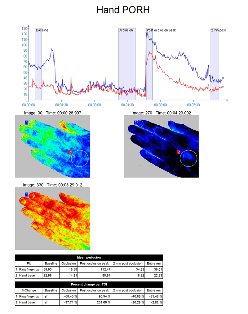
Post-occlusive reactive hyperemia (PORH)
Measure the surge in blood flow that occurs after a temporary prolonged occlusion.
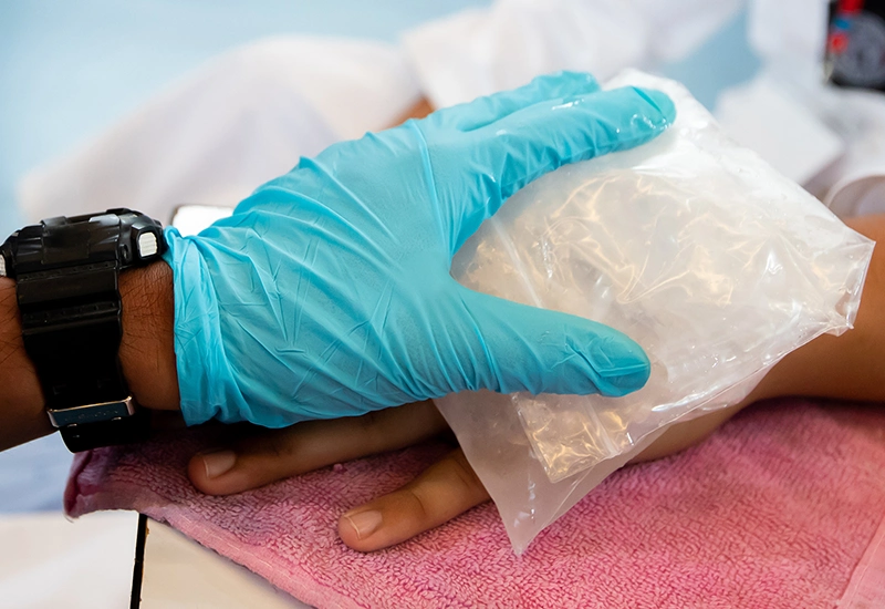
Thermal challenge
Asses how a person responds to heat and cold challenges.
Iontophoresis
Assess a person’s response to medication or other therapeutic agents delivered noninvasively through the skin.
Contact US
Get in touch
If you would like to know more about PeriCam PSI NR, fill out the form and we will put a local representative in touch with you.
LASCA and LSCI
Laser speckle contrast imaging (LSCI) is the innovative technology behind PeriCam PSI, offering non-contact, high-resolution perfusion imaging. The technique was first described by J.D. Briers and S. Webster in 1996 and referred to as laser speckle contrast analysis (LASCA). In later years, the term LSCI gained in popularity and is nowadays the more common term. However, LSCI and LASCA both refer to the same technique, providing precise and reliable perfusion data without the need for contrast agents.
References
- Zötterman, J., Opsomer, D., Farnebo, S., Blondeel, P., Monstrey, S., & Tesselaar, E. (2020). Intraoperative Laser Speckle Contrast Imaging in DIEP Breast Reconstruction: A Prospective Case Series Study. Plastic and reconstructive surgery. Global open, 8(1), e2529. https://doi.org/10.1097/GOX.0000000000002529